{"title":"轻度形式的poc1b相关视网膜营养不良,相对保存锥体系统功能。","authors":"Takaaki Hayashi, Kei Mizobuchi, Shuhei Kameya, Shinji Ueno, Tomokazu Matsuura, Tadashi Nakano","doi":"10.1007/s10633-023-09936-9","DOIUrl":null,"url":null,"abstract":"<p><strong>Purpose: </strong>Biallelic variants in POC1B are rare causes of autosomal recessive cone dystrophy associated with generalized cone system dysfunction. In this report, we describe the clinical characteristics of a Japanese male patient with POC1B-associated retinopathy with relatively preserved cone system function.</p><p><strong>Methods: </strong>We performed whole-exome sequencing (WES) to identify the disease-causing variants and a comprehensive ophthalmic examination, including full-field and multifocal electroretinography (ffERG and mfERG).</p><p><strong>Results: </strong>Our WES analysis identified novel compound heterozygous POC1B variants (p.Arg106Gln and p.Arg452Ter) in the patient. His unaffected mother carried the p.Arg452Ter variant heterozygously. The patient experienced decreased visual acuity in his 50s. At the age of 63, his corrected visual acuity was 20/22 in the right and 20/20 in the left eye. Fundus and fundus autofluorescence images for each eye showed no remarkable finding, except for a subtle hyperautofluorescent spot in the fovea of the left eye. Cross-sectional optical coherence tomography demonstrated blurred but a relatively preserved ellipsoid zone. The ffERG showed that amplitudes of rod and standard-flash responses were within the reference range, whereas the cone and light-adapted 30-Hz flicker amplitudes were close to, or slightly below, the reference range. The mfERG revealed substantially reduced responses with relative preservation of central function.</p><p><strong>Conclusions: </strong>We reported the case of an older patient with POC1B-associated retinopathy, demonstrating late-onset visual decrease, good visual acuity, and relatively preserved cone system function. The disease condition was much milder than previously reported in patients with POC1B-associated retinopathy.</p>","PeriodicalId":11207,"journal":{"name":"Documenta Ophthalmologica","volume":"147 1","pages":"59-70"},"PeriodicalIF":2.6000,"publicationDate":"2023-08-01","publicationTypes":"Journal Article","fieldsOfStudy":null,"isOpenAccess":false,"openAccessPdf":"","citationCount":"0","resultStr":"{\"title\":\"A mild form of POC1B-associated retinal dystrophy with relatively preserved cone system function.\",\"authors\":\"Takaaki Hayashi, Kei Mizobuchi, Shuhei Kameya, Shinji Ueno, Tomokazu Matsuura, Tadashi Nakano\",\"doi\":\"10.1007/s10633-023-09936-9\",\"DOIUrl\":null,\"url\":null,\"abstract\":\"<p><strong>Purpose: </strong>Biallelic variants in POC1B are rare causes of autosomal recessive cone dystrophy associated with generalized cone system dysfunction. In this report, we describe the clinical characteristics of a Japanese male patient with POC1B-associated retinopathy with relatively preserved cone system function.</p><p><strong>Methods: </strong>We performed whole-exome sequencing (WES) to identify the disease-causing variants and a comprehensive ophthalmic examination, including full-field and multifocal electroretinography (ffERG and mfERG).</p><p><strong>Results: </strong>Our WES analysis identified novel compound heterozygous POC1B variants (p.Arg106Gln and p.Arg452Ter) in the patient. His unaffected mother carried the p.Arg452Ter variant heterozygously. The patient experienced decreased visual acuity in his 50s. At the age of 63, his corrected visual acuity was 20/22 in the right and 20/20 in the left eye. Fundus and fundus autofluorescence images for each eye showed no remarkable finding, except for a subtle hyperautofluorescent spot in the fovea of the left eye. Cross-sectional optical coherence tomography demonstrated blurred but a relatively preserved ellipsoid zone. The ffERG showed that amplitudes of rod and standard-flash responses were within the reference range, whereas the cone and light-adapted 30-Hz flicker amplitudes were close to, or slightly below, the reference range. The mfERG revealed substantially reduced responses with relative preservation of central function.</p><p><strong>Conclusions: </strong>We reported the case of an older patient with POC1B-associated retinopathy, demonstrating late-onset visual decrease, good visual acuity, and relatively preserved cone system function. The disease condition was much milder than previously reported in patients with POC1B-associated retinopathy.</p>\",\"PeriodicalId\":11207,\"journal\":{\"name\":\"Documenta Ophthalmologica\",\"volume\":\"147 1\",\"pages\":\"59-70\"},\"PeriodicalIF\":2.6000,\"publicationDate\":\"2023-08-01\",\"publicationTypes\":\"Journal Article\",\"fieldsOfStudy\":null,\"isOpenAccess\":false,\"openAccessPdf\":\"\",\"citationCount\":\"0\",\"resultStr\":null,\"platform\":\"Semanticscholar\",\"paperid\":null,\"PeriodicalName\":\"Documenta Ophthalmologica\",\"FirstCategoryId\":\"3\",\"ListUrlMain\":\"https://doi.org/10.1007/s10633-023-09936-9\",\"RegionNum\":4,\"RegionCategory\":\"医学\",\"ArticlePicture\":[],\"TitleCN\":null,\"AbstractTextCN\":null,\"PMCID\":null,\"EPubDate\":\"\",\"PubModel\":\"\",\"JCR\":\"Q2\",\"JCRName\":\"OPHTHALMOLOGY\",\"Score\":null,\"Total\":0}","platform":"Semanticscholar","paperid":null,"PeriodicalName":"Documenta Ophthalmologica","FirstCategoryId":"3","ListUrlMain":"https://doi.org/10.1007/s10633-023-09936-9","RegionNum":4,"RegionCategory":"医学","ArticlePicture":[],"TitleCN":null,"AbstractTextCN":null,"PMCID":null,"EPubDate":"","PubModel":"","JCR":"Q2","JCRName":"OPHTHALMOLOGY","Score":null,"Total":0}
A mild form of POC1B-associated retinal dystrophy with relatively preserved cone system function.
Purpose: Biallelic variants in POC1B are rare causes of autosomal recessive cone dystrophy associated with generalized cone system dysfunction. In this report, we describe the clinical characteristics of a Japanese male patient with POC1B-associated retinopathy with relatively preserved cone system function.
Methods: We performed whole-exome sequencing (WES) to identify the disease-causing variants and a comprehensive ophthalmic examination, including full-field and multifocal electroretinography (ffERG and mfERG).
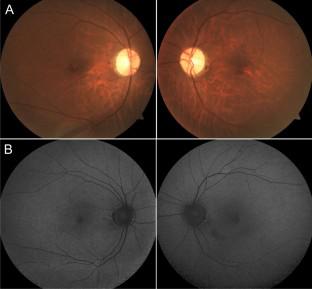
Results: Our WES analysis identified novel compound heterozygous POC1B variants (p.Arg106Gln and p.Arg452Ter) in the patient. His unaffected mother carried the p.Arg452Ter variant heterozygously. The patient experienced decreased visual acuity in his 50s. At the age of 63, his corrected visual acuity was 20/22 in the right and 20/20 in the left eye. Fundus and fundus autofluorescence images for each eye showed no remarkable finding, except for a subtle hyperautofluorescent spot in the fovea of the left eye. Cross-sectional optical coherence tomography demonstrated blurred but a relatively preserved ellipsoid zone. The ffERG showed that amplitudes of rod and standard-flash responses were within the reference range, whereas the cone and light-adapted 30-Hz flicker amplitudes were close to, or slightly below, the reference range. The mfERG revealed substantially reduced responses with relative preservation of central function.
Conclusions: We reported the case of an older patient with POC1B-associated retinopathy, demonstrating late-onset visual decrease, good visual acuity, and relatively preserved cone system function. The disease condition was much milder than previously reported in patients with POC1B-associated retinopathy.
期刊介绍:
Documenta Ophthalmologica is an official publication of the International Society for Clinical Electrophysiology of Vision. The purpose of the journal is to promote the understanding and application of clinical electrophysiology of vision. Documenta Ophthalmologica will publish reviews, research articles, technical notes, brief reports and case studies which inform the readers about basic and clinical sciences related to visual electrodiagnosis and means to improve diagnosis and clinical management of patients using visual electrophysiology. Studies may involve animals or humans. In either case appropriate care must be taken to follow the Declaration of Helsinki for human subject or appropriate humane standards of animal care (e.g., the ARVO standards on Animal Care and Use).

 求助内容:
求助内容: 应助结果提醒方式:


