{"title":"杆状光感受器在早期糖尿病性眼病中的激活和失活。","authors":"J Jason McAnany, Jason C Park","doi":"10.1007/s10633-023-09925-y","DOIUrl":null,"url":null,"abstract":"<p><strong>Purpose: </strong>To infer rod phototransduction activation and deactivation characteristics in diabetics who have mild or no clinically-apparent retinopathy.</p><p><strong>Methods: </strong>Fifteen non-diabetic controls, 15 diabetics with no clinically-apparent diabetic retinopathy (NDR), and 15 diabetics with mild non-proliferative diabetic retinopathy (MDR) participated. Dark-adapted flash electroretinograms (3.2 to 4.4 log scot td-s) were recorded to assess rod activation. The a-waves were fit with a Gaussian model to derive R<sub>mp3</sub> (maximum photoreceptor response amplitude) and S (phototransduction sensitivity). Rod deactivation was assessed with a paired flash paradigm, in which a-waves were measured for two flashes separated by inter-stimulus intervals (ISIs) of 0.125 to 16 s. The ISI needed for the a-wave amplitude of the second flash to recover to 50% of the first flash (t<sub>50</sub>) was determined. The effect of stimulus retinal illuminance on activation and deactivation was evaluated in a subset of control subjects.</p><p><strong>Results: </strong>Analysis of variance indicated that both diabetic groups had significant log S reductions compared to controls (p < 0.001). Mean S was reduced by approximately 49% and 78% for the NDR and MDR groups, respectively. In contrast, log R<sub>mp3</sub> and log t<sub>50</sub> did not differ significantly among the groups (both p > 0.08). Reducing stimulus retinal illuminance significantly reduced S, but did not significantly affect R<sub>max</sub> or t<sub>50</sub>.</p><p><strong>Conclusions: </strong>Only phototransduction sensitivity was abnormal in this sample of diabetic subjects. The normal deactivation kinetics suggests that circulating rod current is normal. These findings begin to constrain possible explanations for abnormal rod function in early diabetic retinal disease.</p>","PeriodicalId":11207,"journal":{"name":"Documenta Ophthalmologica","volume":"146 3","pages":"229-239"},"PeriodicalIF":2.6000,"publicationDate":"2023-06-01","publicationTypes":"Journal Article","fieldsOfStudy":null,"isOpenAccess":false,"openAccessPdf":"","citationCount":"2","resultStr":"{\"title\":\"Rod photoreceptor activation and deactivation in early-stage diabetic eye disease.\",\"authors\":\"J Jason McAnany, Jason C Park\",\"doi\":\"10.1007/s10633-023-09925-y\",\"DOIUrl\":null,\"url\":null,\"abstract\":\"<p><strong>Purpose: </strong>To infer rod phototransduction activation and deactivation characteristics in diabetics who have mild or no clinically-apparent retinopathy.</p><p><strong>Methods: </strong>Fifteen non-diabetic controls, 15 diabetics with no clinically-apparent diabetic retinopathy (NDR), and 15 diabetics with mild non-proliferative diabetic retinopathy (MDR) participated. Dark-adapted flash electroretinograms (3.2 to 4.4 log scot td-s) were recorded to assess rod activation. The a-waves were fit with a Gaussian model to derive R<sub>mp3</sub> (maximum photoreceptor response amplitude) and S (phototransduction sensitivity). Rod deactivation was assessed with a paired flash paradigm, in which a-waves were measured for two flashes separated by inter-stimulus intervals (ISIs) of 0.125 to 16 s. The ISI needed for the a-wave amplitude of the second flash to recover to 50% of the first flash (t<sub>50</sub>) was determined. The effect of stimulus retinal illuminance on activation and deactivation was evaluated in a subset of control subjects.</p><p><strong>Results: </strong>Analysis of variance indicated that both diabetic groups had significant log S reductions compared to controls (p < 0.001). Mean S was reduced by approximately 49% and 78% for the NDR and MDR groups, respectively. In contrast, log R<sub>mp3</sub> and log t<sub>50</sub> did not differ significantly among the groups (both p > 0.08). Reducing stimulus retinal illuminance significantly reduced S, but did not significantly affect R<sub>max</sub> or t<sub>50</sub>.</p><p><strong>Conclusions: </strong>Only phototransduction sensitivity was abnormal in this sample of diabetic subjects. The normal deactivation kinetics suggests that circulating rod current is normal. These findings begin to constrain possible explanations for abnormal rod function in early diabetic retinal disease.</p>\",\"PeriodicalId\":11207,\"journal\":{\"name\":\"Documenta Ophthalmologica\",\"volume\":\"146 3\",\"pages\":\"229-239\"},\"PeriodicalIF\":2.6000,\"publicationDate\":\"2023-06-01\",\"publicationTypes\":\"Journal Article\",\"fieldsOfStudy\":null,\"isOpenAccess\":false,\"openAccessPdf\":\"\",\"citationCount\":\"2\",\"resultStr\":null,\"platform\":\"Semanticscholar\",\"paperid\":null,\"PeriodicalName\":\"Documenta Ophthalmologica\",\"FirstCategoryId\":\"3\",\"ListUrlMain\":\"https://doi.org/10.1007/s10633-023-09925-y\",\"RegionNum\":4,\"RegionCategory\":\"医学\",\"ArticlePicture\":[],\"TitleCN\":null,\"AbstractTextCN\":null,\"PMCID\":null,\"EPubDate\":\"\",\"PubModel\":\"\",\"JCR\":\"Q2\",\"JCRName\":\"OPHTHALMOLOGY\",\"Score\":null,\"Total\":0}","platform":"Semanticscholar","paperid":null,"PeriodicalName":"Documenta Ophthalmologica","FirstCategoryId":"3","ListUrlMain":"https://doi.org/10.1007/s10633-023-09925-y","RegionNum":4,"RegionCategory":"医学","ArticlePicture":[],"TitleCN":null,"AbstractTextCN":null,"PMCID":null,"EPubDate":"","PubModel":"","JCR":"Q2","JCRName":"OPHTHALMOLOGY","Score":null,"Total":0}
Rod photoreceptor activation and deactivation in early-stage diabetic eye disease.
Purpose: To infer rod phototransduction activation and deactivation characteristics in diabetics who have mild or no clinically-apparent retinopathy.
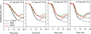
Methods: Fifteen non-diabetic controls, 15 diabetics with no clinically-apparent diabetic retinopathy (NDR), and 15 diabetics with mild non-proliferative diabetic retinopathy (MDR) participated. Dark-adapted flash electroretinograms (3.2 to 4.4 log scot td-s) were recorded to assess rod activation. The a-waves were fit with a Gaussian model to derive Rmp3 (maximum photoreceptor response amplitude) and S (phototransduction sensitivity). Rod deactivation was assessed with a paired flash paradigm, in which a-waves were measured for two flashes separated by inter-stimulus intervals (ISIs) of 0.125 to 16 s. The ISI needed for the a-wave amplitude of the second flash to recover to 50% of the first flash (t50) was determined. The effect of stimulus retinal illuminance on activation and deactivation was evaluated in a subset of control subjects.
Results: Analysis of variance indicated that both diabetic groups had significant log S reductions compared to controls (p < 0.001). Mean S was reduced by approximately 49% and 78% for the NDR and MDR groups, respectively. In contrast, log Rmp3 and log t50 did not differ significantly among the groups (both p > 0.08). Reducing stimulus retinal illuminance significantly reduced S, but did not significantly affect Rmax or t50.
Conclusions: Only phototransduction sensitivity was abnormal in this sample of diabetic subjects. The normal deactivation kinetics suggests that circulating rod current is normal. These findings begin to constrain possible explanations for abnormal rod function in early diabetic retinal disease.
期刊介绍:
Documenta Ophthalmologica is an official publication of the International Society for Clinical Electrophysiology of Vision. The purpose of the journal is to promote the understanding and application of clinical electrophysiology of vision. Documenta Ophthalmologica will publish reviews, research articles, technical notes, brief reports and case studies which inform the readers about basic and clinical sciences related to visual electrodiagnosis and means to improve diagnosis and clinical management of patients using visual electrophysiology. Studies may involve animals or humans. In either case appropriate care must be taken to follow the Declaration of Helsinki for human subject or appropriate humane standards of animal care (e.g., the ARVO standards on Animal Care and Use).

 求助内容:
求助内容: 应助结果提醒方式:


