先天性皮肤肥大细胞增多症被误认为是非意外损伤。
IF 2
4区 医学
Q2 PEDIATRICS
引用次数: 0
摘要
本文章由计算机程序翻译,如有差异,请以英文原文为准。
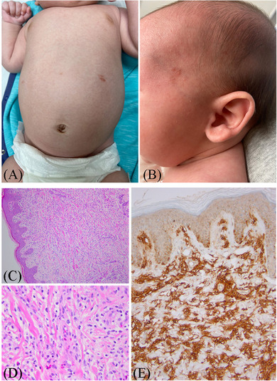
Congenital cutaneous mastocytosis mistaken for non-accidental injury.
求助全文
通过发布文献求助,成功后即可免费获取论文全文。
去求助
来源期刊
Pediatric Investigation
Medicine-Pediatrics, Perinatology and Child Health
CiteScore
3.30
自引率
0.00%
发文量
176
审稿时长
12 weeks
 求助内容:
求助内容: 应助结果提醒方式:


