Alice M Stamatakis;Shanna L Resendez;Kai-Siang Chen;Morgana Favero;Jing Liang-Guallpa;Jonathan J Nassi;Shay Q Neufeld;Koen Visscher;Kunal K Ghosh
{"title":"用于操纵和记录体内大脑活动的微型显微镜","authors":"Alice M Stamatakis;Shanna L Resendez;Kai-Siang Chen;Morgana Favero;Jing Liang-Guallpa;Jonathan J Nassi;Shay Q Neufeld;Koen Visscher;Kunal K Ghosh","doi":"10.1093/jmicro/dfab028","DOIUrl":null,"url":null,"abstract":"Here we describe the development and application of miniature integrated microscopes (miniscopes) paired with microendoscopes that allow for the visualization and manipulation of neural circuits in superficial and subcortical brain regions in freely behaving animals. Over the past decade the miniscope platform has expanded to include simultaneous optogenetic capabilities, electrically-tunable lenses that enable multi-plane imaging, color-corrected optics, and an integrated data acquisition platform that streamlines multimodal experiments. Miniscopes have given researchers an unprecedented ability to monitor hundreds to thousands of genetically-defined neurons from weeks to months in both healthy and diseased animal brains. Sophisticated algorithms that take advantage of constrained matrix factorization allow for background estimation and reliable cell identification, greatly improving the reliability and scalability of source extraction for large imaging datasets. Data generated from miniscopes have empowered researchers to investigate the neural circuit underpinnings of a wide array of behaviors that cannot be studied under head-fixed conditions, such as sleep, reward seeking, learning and memory, social behaviors, and feeding. Importantly, the miniscope has broadened our understanding of how neural circuits can go awry in animal models of progressive neurological disorders, such as Parkinson's disease. Continued miniscope development, including the ability to record from multiple populations of cells simultaneously, along with continued multimodal integration of techniques such as electrophysiology, will allow for deeper understanding into the neural circuits that underlie complex and naturalistic behavior.","PeriodicalId":18515,"journal":{"name":"Microscopy","volume":"70 5","pages":"399-414"},"PeriodicalIF":1.8000,"publicationDate":"2021-10-01","publicationTypes":"Journal Article","fieldsOfStudy":null,"isOpenAccess":false,"openAccessPdf":"https://ftp.ncbi.nlm.nih.gov/pub/pmc/oa_pdf/84/95/dfab028.PMC8491619.pdf","citationCount":"15","resultStr":"{\"title\":\"Miniature microscopes for manipulating and recording in vivo brain activity\",\"authors\":\"Alice M Stamatakis;Shanna L Resendez;Kai-Siang Chen;Morgana Favero;Jing Liang-Guallpa;Jonathan J Nassi;Shay Q Neufeld;Koen Visscher;Kunal K Ghosh\",\"doi\":\"10.1093/jmicro/dfab028\",\"DOIUrl\":null,\"url\":null,\"abstract\":\"Here we describe the development and application of miniature integrated microscopes (miniscopes) paired with microendoscopes that allow for the visualization and manipulation of neural circuits in superficial and subcortical brain regions in freely behaving animals. Over the past decade the miniscope platform has expanded to include simultaneous optogenetic capabilities, electrically-tunable lenses that enable multi-plane imaging, color-corrected optics, and an integrated data acquisition platform that streamlines multimodal experiments. Miniscopes have given researchers an unprecedented ability to monitor hundreds to thousands of genetically-defined neurons from weeks to months in both healthy and diseased animal brains. Sophisticated algorithms that take advantage of constrained matrix factorization allow for background estimation and reliable cell identification, greatly improving the reliability and scalability of source extraction for large imaging datasets. Data generated from miniscopes have empowered researchers to investigate the neural circuit underpinnings of a wide array of behaviors that cannot be studied under head-fixed conditions, such as sleep, reward seeking, learning and memory, social behaviors, and feeding. Importantly, the miniscope has broadened our understanding of how neural circuits can go awry in animal models of progressive neurological disorders, such as Parkinson's disease. Continued miniscope development, including the ability to record from multiple populations of cells simultaneously, along with continued multimodal integration of techniques such as electrophysiology, will allow for deeper understanding into the neural circuits that underlie complex and naturalistic behavior.\",\"PeriodicalId\":18515,\"journal\":{\"name\":\"Microscopy\",\"volume\":\"70 5\",\"pages\":\"399-414\"},\"PeriodicalIF\":1.8000,\"publicationDate\":\"2021-10-01\",\"publicationTypes\":\"Journal Article\",\"fieldsOfStudy\":null,\"isOpenAccess\":false,\"openAccessPdf\":\"https://ftp.ncbi.nlm.nih.gov/pub/pmc/oa_pdf/84/95/dfab028.PMC8491619.pdf\",\"citationCount\":\"15\",\"resultStr\":null,\"platform\":\"Semanticscholar\",\"paperid\":null,\"PeriodicalName\":\"Microscopy\",\"FirstCategoryId\":\"5\",\"ListUrlMain\":\"https://ieeexplore.ieee.org/document/9623671/\",\"RegionNum\":4,\"RegionCategory\":\"工程技术\",\"ArticlePicture\":[],\"TitleCN\":null,\"AbstractTextCN\":null,\"PMCID\":null,\"EPubDate\":\"\",\"PubModel\":\"\",\"JCR\":\"\",\"JCRName\":\"\",\"Score\":null,\"Total\":0}","platform":"Semanticscholar","paperid":null,"PeriodicalName":"Microscopy","FirstCategoryId":"5","ListUrlMain":"https://ieeexplore.ieee.org/document/9623671/","RegionNum":4,"RegionCategory":"工程技术","ArticlePicture":[],"TitleCN":null,"AbstractTextCN":null,"PMCID":null,"EPubDate":"","PubModel":"","JCR":"","JCRName":"","Score":null,"Total":0}
Miniature microscopes for manipulating and recording in vivo brain activity
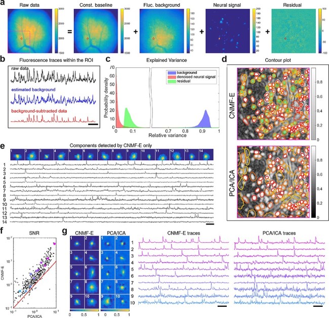
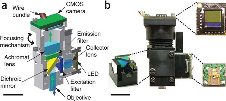
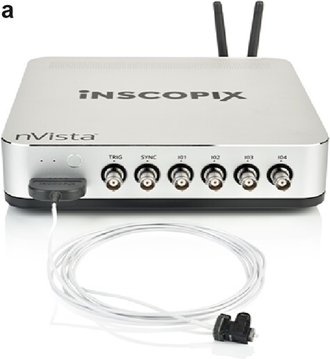
Here we describe the development and application of miniature integrated microscopes (miniscopes) paired with microendoscopes that allow for the visualization and manipulation of neural circuits in superficial and subcortical brain regions in freely behaving animals. Over the past decade the miniscope platform has expanded to include simultaneous optogenetic capabilities, electrically-tunable lenses that enable multi-plane imaging, color-corrected optics, and an integrated data acquisition platform that streamlines multimodal experiments. Miniscopes have given researchers an unprecedented ability to monitor hundreds to thousands of genetically-defined neurons from weeks to months in both healthy and diseased animal brains. Sophisticated algorithms that take advantage of constrained matrix factorization allow for background estimation and reliable cell identification, greatly improving the reliability and scalability of source extraction for large imaging datasets. Data generated from miniscopes have empowered researchers to investigate the neural circuit underpinnings of a wide array of behaviors that cannot be studied under head-fixed conditions, such as sleep, reward seeking, learning and memory, social behaviors, and feeding. Importantly, the miniscope has broadened our understanding of how neural circuits can go awry in animal models of progressive neurological disorders, such as Parkinson's disease. Continued miniscope development, including the ability to record from multiple populations of cells simultaneously, along with continued multimodal integration of techniques such as electrophysiology, will allow for deeper understanding into the neural circuits that underlie complex and naturalistic behavior.
期刊介绍:
Microscopy, previously Journal of Electron Microscopy, promotes research combined with any type of microscopy techniques, applied in life and material sciences. Microscopy is the official journal of the Japanese Society of Microscopy.
 求助内容:
求助内容: 应助结果提醒方式:


