利用FIB-SEM研究细胞器动力学在光镜活体成像和电子显微镜三维结构之间的相关性

IF 1.8 4区 工程技术
Microscopy Pub Date : 2020-11-01 DOI:10.1093/jmicro/dfaa071
Keisuke Ohta;Shingo Hirashima;Yoshihiro Miyazono;Akinobu Togo;Kei-ichiro Nakamura
{"title":"利用FIB-SEM研究细胞器动力学在光镜活体成像和电子显微镜三维结构之间的相关性","authors":"Keisuke Ohta;Shingo Hirashima;Yoshihiro Miyazono;Akinobu Togo;Kei-ichiro Nakamura","doi":"10.1093/jmicro/dfaa071","DOIUrl":null,"url":null,"abstract":"Correlative light and electron microscopy (CLEM) methods combined with live imaging can be applied to understand the dynamics of organelles. Although recent advances in cell biology and light microscopy have helped in visualizing the details of organelle activities, observing their ultrastructure or organization of surrounding microenvironments is a challenging task. Therefore, CLEM, which allows us to observe the same area as an optical microscope with an electron microscope, has become a key technique in cell biology. Unfortunately, most CLEM methods have technical drawbacks, and many researchers face difficulties in applying CLEM methods. Here, we propose a live three-dimensional CLEM method, combined with a three-dimensional reconstruction technique using focused ion beam scanning electron microscopy tomography, as a solution to such technical barriers. We review our method, the associated technical limitations and the options considered to perform live CLEM.","PeriodicalId":18515,"journal":{"name":"Microscopy","volume":null,"pages":null},"PeriodicalIF":1.8000,"publicationDate":"2020-11-01","publicationTypes":"Journal Article","fieldsOfStudy":null,"isOpenAccess":false,"openAccessPdf":"https://sci-hub-pdf.com/10.1093/jmicro/dfaa071","citationCount":"7","resultStr":"{\"title\":\"Correlation of organelle dynamics between light microscopic live imaging and electron microscopic 3D architecture using FIB-SEM\",\"authors\":\"Keisuke Ohta;Shingo Hirashima;Yoshihiro Miyazono;Akinobu Togo;Kei-ichiro Nakamura\",\"doi\":\"10.1093/jmicro/dfaa071\",\"DOIUrl\":null,\"url\":null,\"abstract\":\"Correlative light and electron microscopy (CLEM) methods combined with live imaging can be applied to understand the dynamics of organelles. Although recent advances in cell biology and light microscopy have helped in visualizing the details of organelle activities, observing their ultrastructure or organization of surrounding microenvironments is a challenging task. Therefore, CLEM, which allows us to observe the same area as an optical microscope with an electron microscope, has become a key technique in cell biology. Unfortunately, most CLEM methods have technical drawbacks, and many researchers face difficulties in applying CLEM methods. Here, we propose a live three-dimensional CLEM method, combined with a three-dimensional reconstruction technique using focused ion beam scanning electron microscopy tomography, as a solution to such technical barriers. We review our method, the associated technical limitations and the options considered to perform live CLEM.\",\"PeriodicalId\":18515,\"journal\":{\"name\":\"Microscopy\",\"volume\":null,\"pages\":null},\"PeriodicalIF\":1.8000,\"publicationDate\":\"2020-11-01\",\"publicationTypes\":\"Journal Article\",\"fieldsOfStudy\":null,\"isOpenAccess\":false,\"openAccessPdf\":\"https://sci-hub-pdf.com/10.1093/jmicro/dfaa071\",\"citationCount\":\"7\",\"resultStr\":null,\"platform\":\"Semanticscholar\",\"paperid\":null,\"PeriodicalName\":\"Microscopy\",\"FirstCategoryId\":\"5\",\"ListUrlMain\":\"https://ieeexplore.ieee.org/document/9433138/\",\"RegionNum\":4,\"RegionCategory\":\"工程技术\",\"ArticlePicture\":[],\"TitleCN\":null,\"AbstractTextCN\":null,\"PMCID\":null,\"EPubDate\":\"\",\"PubModel\":\"\",\"JCR\":\"\",\"JCRName\":\"\",\"Score\":null,\"Total\":0}","platform":"Semanticscholar","paperid":null,"PeriodicalName":"Microscopy","FirstCategoryId":"5","ListUrlMain":"https://ieeexplore.ieee.org/document/9433138/","RegionNum":4,"RegionCategory":"工程技术","ArticlePicture":[],"TitleCN":null,"AbstractTextCN":null,"PMCID":null,"EPubDate":"","PubModel":"","JCR":"","JCRName":"","Score":null,"Total":0}
引用次数: 7

摘要

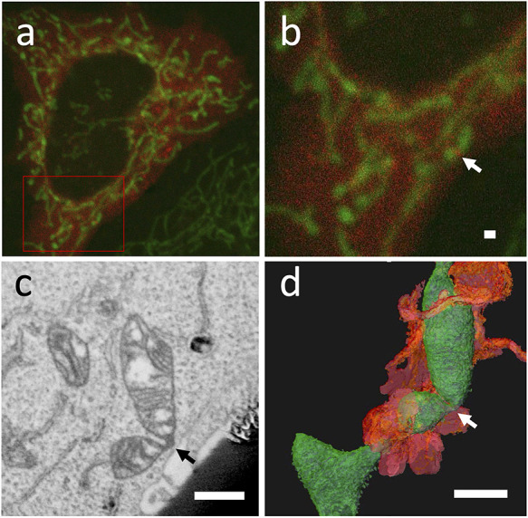
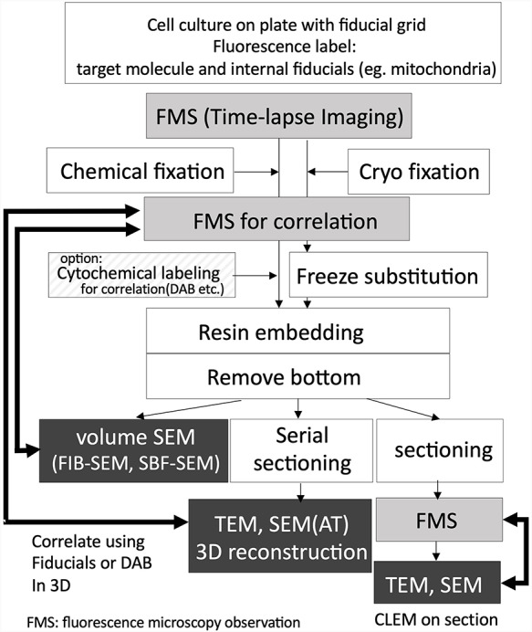
相关的光学和电子显微镜(CLEM)方法结合实时成像可以用于了解细胞器的动力学。尽管细胞生物学和光学显微镜的最新进展有助于可视化细胞器活动的细节,但观察细胞器的超微结构或周围微环境的组织是一项具有挑战性的任务。因此,CLEM使我们能够用电子显微镜观察与光学显微镜相同的区域,已成为细胞生物学中的一项关键技术。不幸的是,大多数CLEM方法都有技术缺陷,许多研究人员在应用CLEM方法时面临困难。在这里,我们提出了一种实时三维CLEM方法,结合使用聚焦离子束扫描电子显微镜断层扫描的三维重建技术,作为解决这些技术障碍的方法。我们回顾了我们的方法、相关的技术限制以及执行现场CLEM所考虑的选项。
本文章由计算机程序翻译,如有差异,请以英文原文为准。

Correlation of organelle dynamics between light microscopic live imaging and electron microscopic 3D architecture using FIB-SEM

Correlation of organelle dynamics between light microscopic live imaging and electron microscopic 3D architecture using FIB-SEM

Correlation of organelle dynamics between light microscopic live imaging and electron microscopic 3D architecture using FIB-SEM

Correlation of organelle dynamics between light microscopic live imaging and electron microscopic 3D architecture using FIB-SEM
Correlative light and electron microscopy (CLEM) methods combined with live imaging can be applied to understand the dynamics of organelles. Although recent advances in cell biology and light microscopy have helped in visualizing the details of organelle activities, observing their ultrastructure or organization of surrounding microenvironments is a challenging task. Therefore, CLEM, which allows us to observe the same area as an optical microscope with an electron microscope, has become a key technique in cell biology. Unfortunately, most CLEM methods have technical drawbacks, and many researchers face difficulties in applying CLEM methods. Here, we propose a live three-dimensional CLEM method, combined with a three-dimensional reconstruction technique using focused ion beam scanning electron microscopy tomography, as a solution to such technical barriers. We review our method, the associated technical limitations and the options considered to perform live CLEM.
求助全文
通过发布文献求助,成功后即可免费获取论文全文。 去求助
来源期刊
Microscopy
Microscopy 工程技术-显微镜技术
自引率
11.10%
发文量
0
审稿时长
>12 weeks
期刊介绍: Microscopy, previously Journal of Electron Microscopy, promotes research combined with any type of microscopy techniques, applied in life and material sciences. Microscopy is the official journal of the Japanese Society of Microscopy.
×
引用
GB/T 7714-2015
复制
MLA
复制
APA
复制
导出至
BibTeX EndNote RefMan NoteFirst NoteExpress
×
提示
您的信息不完整,为了账户安全,请先补充。
现在去补充
×
提示
您因"违规操作"
具体请查看互助需知
我知道了
×
提示
确定
请完成安全验证×
copy
已复制链接
快去分享给好友吧!
我知道了
右上角分享
点击右上角分享
0
联系我们:info@booksci.cn Book学术提供免费学术资源搜索服务,方便国内外学者检索中英文文献。致力于提供最便捷和优质的服务体验。 Copyright © 2023 布克学术 All rights reserved.
京ICP备2023020795号-1
ghs 京公网安备 11010802042870号
Book学术文献互助
Book学术文献互助群
群 号:481959085
Book学术官方微信