{"title":"长春胺减轻甲氨蝶呤诱导的肝纤维化模型。","authors":"Yonca Yılmaz Ürün, Gürkan Güner, Ejder Saylav Bora, Ayşe Buket Taşkın, Muslih Ürün, Oytun Erbaş","doi":"10.5152/tjg.2025.24716","DOIUrl":null,"url":null,"abstract":"<p><p>Background/Aims: Liver fibrosis is linked to higher rates of death and disease. This study examined the hepatoprotective properties of vincamine and its potential therapeutic application in treating liver damage caused by methotrexate in rats. Materials and Methods: Thirty male Wistar albino rats, with weights ranging from 150 to 200 g and ages between 10 and 12 weeks, were included in the study. A total of 10 rats were selected to serve as the control group, receiving no medication. A group of 20 rats was given a single intraperitoneal dose of 20 mg/kg methotrexate in order to cause liver damage. Subsequently, the participants were randomly allocated into 2 cohorts and administered either 1 mL/kg/day tap water or 50 mg/kg/day vincamine orally through gavage on a daily basis for a duration of 10 days. Following the completion of the treatment period, the animals were euthanized and their livers were examined histologically. Furthermore, the levels of plasma galectin-3 (gal-3), cytokeratin 18, malondialdehyde (MDA), alanine transaminase (ALT), liver MDA, and transforming growth factor beta (TGF-β) levels were evaluated. Results: Treatment with vincamine resulted in a significant decrease in plasma gal-3, cytokeratin, MDA, and ALT levels and liver MDA and TGF-β levels compared to the methotrexate and saline group. Vincamine treatment effectively protected against liver injury, and histopathological examination of the livers confirmed these results. Conclusion: This study demonstrates that vincamine alleviates methotrexate-induced liver toxicity via exhibiting antioxidant, antiinflammatory, and anti-fibrotic activities and improved liver functionally, biochemically, and histopathologically.</p>","PeriodicalId":51205,"journal":{"name":"Turkish Journal of Gastroenterology","volume":"36 10","pages":"641-648"},"PeriodicalIF":1.6000,"publicationDate":"2025-06-23","publicationTypes":"Journal Article","fieldsOfStudy":null,"isOpenAccess":false,"openAccessPdf":"https://www.ncbi.nlm.nih.gov/pmc/articles/PMC12520132/pdf/","citationCount":"0","resultStr":"{\"title\":\"Vincamine Mitigates Methotrexate-Induced Liver Fibrosis Model.\",\"authors\":\"Yonca Yılmaz Ürün, Gürkan Güner, Ejder Saylav Bora, Ayşe Buket Taşkın, Muslih Ürün, Oytun Erbaş\",\"doi\":\"10.5152/tjg.2025.24716\",\"DOIUrl\":null,\"url\":null,\"abstract\":\"<p><p>Background/Aims: Liver fibrosis is linked to higher rates of death and disease. This study examined the hepatoprotective properties of vincamine and its potential therapeutic application in treating liver damage caused by methotrexate in rats. Materials and Methods: Thirty male Wistar albino rats, with weights ranging from 150 to 200 g and ages between 10 and 12 weeks, were included in the study. A total of 10 rats were selected to serve as the control group, receiving no medication. A group of 20 rats was given a single intraperitoneal dose of 20 mg/kg methotrexate in order to cause liver damage. Subsequently, the participants were randomly allocated into 2 cohorts and administered either 1 mL/kg/day tap water or 50 mg/kg/day vincamine orally through gavage on a daily basis for a duration of 10 days. Following the completion of the treatment period, the animals were euthanized and their livers were examined histologically. Furthermore, the levels of plasma galectin-3 (gal-3), cytokeratin 18, malondialdehyde (MDA), alanine transaminase (ALT), liver MDA, and transforming growth factor beta (TGF-β) levels were evaluated. Results: Treatment with vincamine resulted in a significant decrease in plasma gal-3, cytokeratin, MDA, and ALT levels and liver MDA and TGF-β levels compared to the methotrexate and saline group. Vincamine treatment effectively protected against liver injury, and histopathological examination of the livers confirmed these results. Conclusion: This study demonstrates that vincamine alleviates methotrexate-induced liver toxicity via exhibiting antioxidant, antiinflammatory, and anti-fibrotic activities and improved liver functionally, biochemically, and histopathologically.</p>\",\"PeriodicalId\":51205,\"journal\":{\"name\":\"Turkish Journal of Gastroenterology\",\"volume\":\"36 10\",\"pages\":\"641-648\"},\"PeriodicalIF\":1.6000,\"publicationDate\":\"2025-06-23\",\"publicationTypes\":\"Journal Article\",\"fieldsOfStudy\":null,\"isOpenAccess\":false,\"openAccessPdf\":\"https://www.ncbi.nlm.nih.gov/pmc/articles/PMC12520132/pdf/\",\"citationCount\":\"0\",\"resultStr\":null,\"platform\":\"Semanticscholar\",\"paperid\":null,\"PeriodicalName\":\"Turkish Journal of Gastroenterology\",\"FirstCategoryId\":\"3\",\"ListUrlMain\":\"https://doi.org/10.5152/tjg.2025.24716\",\"RegionNum\":4,\"RegionCategory\":\"医学\",\"ArticlePicture\":[],\"TitleCN\":null,\"AbstractTextCN\":null,\"PMCID\":null,\"EPubDate\":\"\",\"PubModel\":\"\",\"JCR\":\"Q4\",\"JCRName\":\"GASTROENTEROLOGY & HEPATOLOGY\",\"Score\":null,\"Total\":0}","platform":"Semanticscholar","paperid":null,"PeriodicalName":"Turkish Journal of Gastroenterology","FirstCategoryId":"3","ListUrlMain":"https://doi.org/10.5152/tjg.2025.24716","RegionNum":4,"RegionCategory":"医学","ArticlePicture":[],"TitleCN":null,"AbstractTextCN":null,"PMCID":null,"EPubDate":"","PubModel":"","JCR":"Q4","JCRName":"GASTROENTEROLOGY & HEPATOLOGY","Score":null,"Total":0}
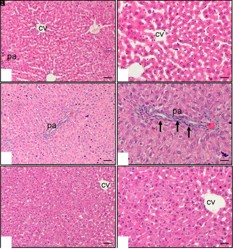
Background/Aims: Liver fibrosis is linked to higher rates of death and disease. This study examined the hepatoprotective properties of vincamine and its potential therapeutic application in treating liver damage caused by methotrexate in rats. Materials and Methods: Thirty male Wistar albino rats, with weights ranging from 150 to 200 g and ages between 10 and 12 weeks, were included in the study. A total of 10 rats were selected to serve as the control group, receiving no medication. A group of 20 rats was given a single intraperitoneal dose of 20 mg/kg methotrexate in order to cause liver damage. Subsequently, the participants were randomly allocated into 2 cohorts and administered either 1 mL/kg/day tap water or 50 mg/kg/day vincamine orally through gavage on a daily basis for a duration of 10 days. Following the completion of the treatment period, the animals were euthanized and their livers were examined histologically. Furthermore, the levels of plasma galectin-3 (gal-3), cytokeratin 18, malondialdehyde (MDA), alanine transaminase (ALT), liver MDA, and transforming growth factor beta (TGF-β) levels were evaluated. Results: Treatment with vincamine resulted in a significant decrease in plasma gal-3, cytokeratin, MDA, and ALT levels and liver MDA and TGF-β levels compared to the methotrexate and saline group. Vincamine treatment effectively protected against liver injury, and histopathological examination of the livers confirmed these results. Conclusion: This study demonstrates that vincamine alleviates methotrexate-induced liver toxicity via exhibiting antioxidant, antiinflammatory, and anti-fibrotic activities and improved liver functionally, biochemically, and histopathologically.
期刊介绍:
The Turkish Journal of Gastroenterology (Turk J Gastroenterol) is the double-blind peer-reviewed, open access, international publication organ of the Turkish Society of Gastroenterology. The journal is a bimonthly publication, published on January, March, May, July, September, November and its publication language is English.
The Turkish Journal of Gastroenterology aims to publish international at the highest clinical and scientific level on original issues of gastroenterology and hepatology. The journal publishes original papers, review articles, case reports and letters to the editor on clinical and experimental gastroenterology and hepatology.

 求助内容:
求助内容: 应助结果提醒方式:


