Ming Wang, Abdukahar Kiram, Jie Li, Yunlong Xu, Jingtan Hu, Xiaodong Qin, Yu Wang, Jun Qiao, Benlong Shi, Saihu Mao, Zezhang Zhu, Yong Qiu, Zhen Liu
{"title":"椎旁肌减少症对退行性后凸矢状位失衡的影响。","authors":"Ming Wang, Abdukahar Kiram, Jie Li, Yunlong Xu, Jingtan Hu, Xiaodong Qin, Yu Wang, Jun Qiao, Benlong Shi, Saihu Mao, Zezhang Zhu, Yong Qiu, Zhen Liu","doi":"10.14245/ns.2550436.218","DOIUrl":null,"url":null,"abstract":"<p><strong>Objective: </strong>To investigate the correlation between paraspinal sarcopenia (PS) and sagittal imbalance (SI) in degenerative kyphosis (DK), and to explore the correlation between paraspinal muscle (PSM) function loss and morphology change in DK.</p><p><strong>Methods: </strong>One hundred thirty-eight patients with DK and 204 with lumbar spinal stenosis (LSS) were enrolled. The spinopelvic parameters and sagittal vertical axis (SVA) were measured. Patients were divided into the sagittal balance (SB, SVA ≤ 5 cm, n = 61) and SI (SVA > 5 cm, n = 77) groups. Sagittal balanced LSS patients were served as control group. PSM function was evaluated by measuring the maximal voluntary exertion (MVE) and endurance time (ET). Magnetic resonance imaging-derived cross-sectional area (CSA) and fat infiltration rate (FI%) of PSM at T10-L5 were normalized to intervertebral disc CSA. Psoas CSA and FI% were calculated at L3-4 disc level. The correlation assessment using Spearman rank correlation coefficient and multiple linear regression. Logistic regression was used to identify the risk factors of SI.</p><p><strong>Results: </strong>Significantly lower ET, MVE, relative CSA (rCSA) and higher rFI% was found in the SI group than in the SB and control. The PS were correlated with spinopelvic parameters and regional kyphosis, while lack of correlation was found between the rFI% and MVE. Logistic regression and Youden index analysis showed ET < 15.5 seconds, MVE < 1.3 N/kg, and rCSA (L1-5) atrophy to be potential risk factors for SI in DK.</p><p><strong>Conclusion: </strong>DK patients with SI demonstrate acerbated PS that indicated by significant PSM dysfunction and morphological alterations. We highlight the significance of PSM combined evaluation and revealed that PS plays an indispensable role in the progression of SI, providing novel insights into the underlying sagittal compensatory mechanisms.</p>","PeriodicalId":19269,"journal":{"name":"Neurospine","volume":"22 3","pages":"680-691"},"PeriodicalIF":3.6000,"publicationDate":"2025-09-01","publicationTypes":"Journal Article","fieldsOfStudy":null,"isOpenAccess":false,"openAccessPdf":"https://www.ncbi.nlm.nih.gov/pmc/articles/PMC12518903/pdf/","citationCount":"0","resultStr":"{\"title\":\"The Contribution of Paraspinal Sarcopenia on Sagittal Imbalance in Degenerative Kyphosis.\",\"authors\":\"Ming Wang, Abdukahar Kiram, Jie Li, Yunlong Xu, Jingtan Hu, Xiaodong Qin, Yu Wang, Jun Qiao, Benlong Shi, Saihu Mao, Zezhang Zhu, Yong Qiu, Zhen Liu\",\"doi\":\"10.14245/ns.2550436.218\",\"DOIUrl\":null,\"url\":null,\"abstract\":\"<p><strong>Objective: </strong>To investigate the correlation between paraspinal sarcopenia (PS) and sagittal imbalance (SI) in degenerative kyphosis (DK), and to explore the correlation between paraspinal muscle (PSM) function loss and morphology change in DK.</p><p><strong>Methods: </strong>One hundred thirty-eight patients with DK and 204 with lumbar spinal stenosis (LSS) were enrolled. The spinopelvic parameters and sagittal vertical axis (SVA) were measured. Patients were divided into the sagittal balance (SB, SVA ≤ 5 cm, n = 61) and SI (SVA > 5 cm, n = 77) groups. Sagittal balanced LSS patients were served as control group. PSM function was evaluated by measuring the maximal voluntary exertion (MVE) and endurance time (ET). Magnetic resonance imaging-derived cross-sectional area (CSA) and fat infiltration rate (FI%) of PSM at T10-L5 were normalized to intervertebral disc CSA. Psoas CSA and FI% were calculated at L3-4 disc level. The correlation assessment using Spearman rank correlation coefficient and multiple linear regression. Logistic regression was used to identify the risk factors of SI.</p><p><strong>Results: </strong>Significantly lower ET, MVE, relative CSA (rCSA) and higher rFI% was found in the SI group than in the SB and control. The PS were correlated with spinopelvic parameters and regional kyphosis, while lack of correlation was found between the rFI% and MVE. Logistic regression and Youden index analysis showed ET < 15.5 seconds, MVE < 1.3 N/kg, and rCSA (L1-5) atrophy to be potential risk factors for SI in DK.</p><p><strong>Conclusion: </strong>DK patients with SI demonstrate acerbated PS that indicated by significant PSM dysfunction and morphological alterations. We highlight the significance of PSM combined evaluation and revealed that PS plays an indispensable role in the progression of SI, providing novel insights into the underlying sagittal compensatory mechanisms.</p>\",\"PeriodicalId\":19269,\"journal\":{\"name\":\"Neurospine\",\"volume\":\"22 3\",\"pages\":\"680-691\"},\"PeriodicalIF\":3.6000,\"publicationDate\":\"2025-09-01\",\"publicationTypes\":\"Journal Article\",\"fieldsOfStudy\":null,\"isOpenAccess\":false,\"openAccessPdf\":\"https://www.ncbi.nlm.nih.gov/pmc/articles/PMC12518903/pdf/\",\"citationCount\":\"0\",\"resultStr\":null,\"platform\":\"Semanticscholar\",\"paperid\":null,\"PeriodicalName\":\"Neurospine\",\"FirstCategoryId\":\"3\",\"ListUrlMain\":\"https://doi.org/10.14245/ns.2550436.218\",\"RegionNum\":2,\"RegionCategory\":\"医学\",\"ArticlePicture\":[],\"TitleCN\":null,\"AbstractTextCN\":null,\"PMCID\":null,\"EPubDate\":\"2025/9/30 0:00:00\",\"PubModel\":\"Epub\",\"JCR\":\"Q1\",\"JCRName\":\"CLINICAL NEUROLOGY\",\"Score\":null,\"Total\":0}","platform":"Semanticscholar","paperid":null,"PeriodicalName":"Neurospine","FirstCategoryId":"3","ListUrlMain":"https://doi.org/10.14245/ns.2550436.218","RegionNum":2,"RegionCategory":"医学","ArticlePicture":[],"TitleCN":null,"AbstractTextCN":null,"PMCID":null,"EPubDate":"2025/9/30 0:00:00","PubModel":"Epub","JCR":"Q1","JCRName":"CLINICAL NEUROLOGY","Score":null,"Total":0}
引用次数: 0
摘要
目的:探讨退行性后凸症(DK)椎旁肌减少症(PS)与矢状面失衡(SI)的相关性,探讨DK椎旁肌(PSM)功能丧失与形态学改变的相关性。方法:纳入138例DK患者和204例腰椎管狭窄(LSS)患者。测量脊柱骨盆参数和矢状垂直轴(SVA)。将患者分为矢状平衡组(SVA≤5 cm, n = 61)和矢状平衡组(SVA≤5 cm, n = 77)。矢状平衡型LSS患者作为对照组。通过测量最大自主用力(MVE)和耐力时间(ET)评价PSM功能。将T10-L5 PSM的磁共振成像衍生横截面积(CSA)和脂肪浸润率(FI%)归一化为椎间盘CSA。在L3-4椎间盘水平计算腰椎间盘CSA和FI%。相关性评价采用Spearman秩相关系数和多元线性回归。采用Logistic回归分析确定SI的危险因素。结果:SI组ET、MVE、相对CSA (rCSA)明显低于SB组和对照组,rFI%明显高于对照组。PS与脊柱骨盆参数和局部后凸相关,而rFI%与MVE之间缺乏相关性。Logistic回归和约登指数分析显示,ET < 15.5秒、MVE < 1.3 N/kg、rCSA (L1-5)萎缩是DK发生SI的潜在危险因素。结论:DK合并SI患者PS加重,表现为明显的PSM功能障碍和形态改变。我们强调了PSM联合评估的重要性,并揭示了PS在SI的进展中起着不可或缺的作用,为潜在的矢状面代偿机制提供了新的见解。
The Contribution of Paraspinal Sarcopenia on Sagittal Imbalance in Degenerative Kyphosis.
Objective: To investigate the correlation between paraspinal sarcopenia (PS) and sagittal imbalance (SI) in degenerative kyphosis (DK), and to explore the correlation between paraspinal muscle (PSM) function loss and morphology change in DK.
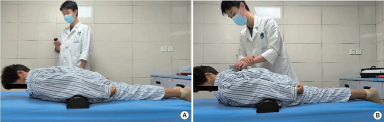
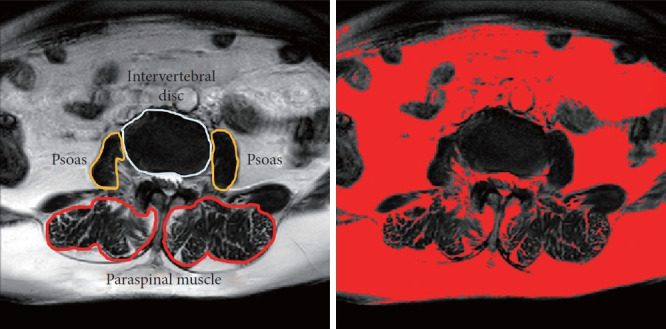
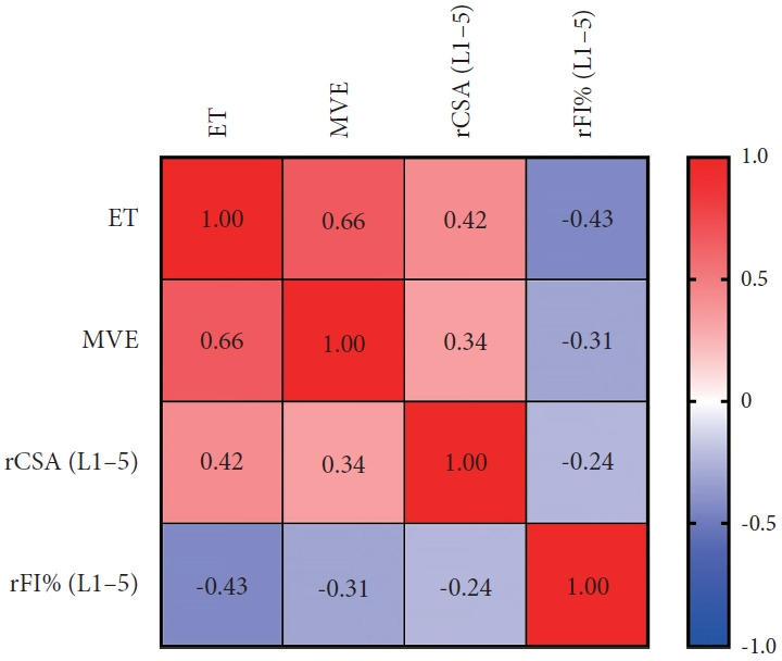
Methods: One hundred thirty-eight patients with DK and 204 with lumbar spinal stenosis (LSS) were enrolled. The spinopelvic parameters and sagittal vertical axis (SVA) were measured. Patients were divided into the sagittal balance (SB, SVA ≤ 5 cm, n = 61) and SI (SVA > 5 cm, n = 77) groups. Sagittal balanced LSS patients were served as control group. PSM function was evaluated by measuring the maximal voluntary exertion (MVE) and endurance time (ET). Magnetic resonance imaging-derived cross-sectional area (CSA) and fat infiltration rate (FI%) of PSM at T10-L5 were normalized to intervertebral disc CSA. Psoas CSA and FI% were calculated at L3-4 disc level. The correlation assessment using Spearman rank correlation coefficient and multiple linear regression. Logistic regression was used to identify the risk factors of SI.
Results: Significantly lower ET, MVE, relative CSA (rCSA) and higher rFI% was found in the SI group than in the SB and control. The PS were correlated with spinopelvic parameters and regional kyphosis, while lack of correlation was found between the rFI% and MVE. Logistic regression and Youden index analysis showed ET < 15.5 seconds, MVE < 1.3 N/kg, and rCSA (L1-5) atrophy to be potential risk factors for SI in DK.
Conclusion: DK patients with SI demonstrate acerbated PS that indicated by significant PSM dysfunction and morphological alterations. We highlight the significance of PSM combined evaluation and revealed that PS plays an indispensable role in the progression of SI, providing novel insights into the underlying sagittal compensatory mechanisms.
 求助内容:
求助内容: 应助结果提醒方式:


