Gi Cheol Park, Soo-Young Bang, Ji Min Kim, Sung-Chan Shin, Yong-Il Cheon, Hanaro Park, Sunghwan Suh, Jung Hwan Cho, Eui-Suk Sung, Minhyung Lee, Jin-Choon Lee, Byung-Joo Lee
{"title":"铁他汀-1通过抑制有丝分裂驱动的铁下垂来预防去卵巢大鼠唾液腺功能障碍。","authors":"Gi Cheol Park, Soo-Young Bang, Ji Min Kim, Sung-Chan Shin, Yong-Il Cheon, Hanaro Park, Sunghwan Suh, Jung Hwan Cho, Eui-Suk Sung, Minhyung Lee, Jin-Choon Lee, Byung-Joo Lee","doi":"10.3390/antiox14091058","DOIUrl":null,"url":null,"abstract":"<p><p>Salivary gland dysfunction is a common but underexplored complication of menopause that contributes to oral dryness, dysphagia, and increased risk of infection. Although ferroptosis, a form of regulated necrotic cell death driven by iron-dependent lipid peroxidation, has recently been implicated in postmenopausal tissue degeneration, its regulatory mechanisms in salivary glands remain unclear. In this study, we investigated the roles of mitochondrial dysfunction and mitophagy in driving ferroptosis-induced salivary gland injury in an ovariectomized (OVX) rat model of estrogen deficiency. OVX rats exhibited elevated markers of oxidative stress, lipid accumulation, and iron overload, and suppression of GPX4 activity in the salivary glands, consistent with ferroptotic activation. These changes were accompanied by impaired mitochondrial dynamics (MFN1 and OPA1), decreased expression of mitochondrial antioxidant regulators (PGC-1α, SOD, and catalase), and upregulation of mitophagy-related genes (<i>PINK1</i>, <i>ULK1</i>, <i>Rab9</i>, and <i>LC3B</i>), as well as LAMP, a lysosomal marker involved in autophagosome-lysosome fusion, while ferritinophagy (NCOA4) remained unchanged. Early administration of ferrostatin-1 effectively suppressed these pathological changes, preserving both glandular structure and function, as evidenced by the restored AQP5 and AMY2A expression. Collectively, our findings reveal that ferroptosis in estrogen-deficient salivary glands is regulated by mitochondrial instability and aberrant mitophagy, and ferrostatin-1 mitigates this cascade through multi-level mitochondrial protection. These results highlight ferrostatin-1 as a promising preventive agent against menopause-associated salivary gland dysfunction, with broader implications for organ-specific ferroptosis modulation.</p>","PeriodicalId":7984,"journal":{"name":"Antioxidants","volume":"14 9","pages":""},"PeriodicalIF":6.6000,"publicationDate":"2025-08-28","publicationTypes":"Journal Article","fieldsOfStudy":null,"isOpenAccess":false,"openAccessPdf":"https://www.ncbi.nlm.nih.gov/pmc/articles/PMC12466376/pdf/","citationCount":"0","resultStr":"{\"title\":\"Ferrostatin-1 Prevents Salivary Gland Dysfunction in an Ovariectomized Rat Model by Suppressing Mitophagy-Driven Ferroptosis.\",\"authors\":\"Gi Cheol Park, Soo-Young Bang, Ji Min Kim, Sung-Chan Shin, Yong-Il Cheon, Hanaro Park, Sunghwan Suh, Jung Hwan Cho, Eui-Suk Sung, Minhyung Lee, Jin-Choon Lee, Byung-Joo Lee\",\"doi\":\"10.3390/antiox14091058\",\"DOIUrl\":null,\"url\":null,\"abstract\":\"<p><p>Salivary gland dysfunction is a common but underexplored complication of menopause that contributes to oral dryness, dysphagia, and increased risk of infection. Although ferroptosis, a form of regulated necrotic cell death driven by iron-dependent lipid peroxidation, has recently been implicated in postmenopausal tissue degeneration, its regulatory mechanisms in salivary glands remain unclear. In this study, we investigated the roles of mitochondrial dysfunction and mitophagy in driving ferroptosis-induced salivary gland injury in an ovariectomized (OVX) rat model of estrogen deficiency. OVX rats exhibited elevated markers of oxidative stress, lipid accumulation, and iron overload, and suppression of GPX4 activity in the salivary glands, consistent with ferroptotic activation. These changes were accompanied by impaired mitochondrial dynamics (MFN1 and OPA1), decreased expression of mitochondrial antioxidant regulators (PGC-1α, SOD, and catalase), and upregulation of mitophagy-related genes (<i>PINK1</i>, <i>ULK1</i>, <i>Rab9</i>, and <i>LC3B</i>), as well as LAMP, a lysosomal marker involved in autophagosome-lysosome fusion, while ferritinophagy (NCOA4) remained unchanged. Early administration of ferrostatin-1 effectively suppressed these pathological changes, preserving both glandular structure and function, as evidenced by the restored AQP5 and AMY2A expression. Collectively, our findings reveal that ferroptosis in estrogen-deficient salivary glands is regulated by mitochondrial instability and aberrant mitophagy, and ferrostatin-1 mitigates this cascade through multi-level mitochondrial protection. These results highlight ferrostatin-1 as a promising preventive agent against menopause-associated salivary gland dysfunction, with broader implications for organ-specific ferroptosis modulation.</p>\",\"PeriodicalId\":7984,\"journal\":{\"name\":\"Antioxidants\",\"volume\":\"14 9\",\"pages\":\"\"},\"PeriodicalIF\":6.6000,\"publicationDate\":\"2025-08-28\",\"publicationTypes\":\"Journal Article\",\"fieldsOfStudy\":null,\"isOpenAccess\":false,\"openAccessPdf\":\"https://www.ncbi.nlm.nih.gov/pmc/articles/PMC12466376/pdf/\",\"citationCount\":\"0\",\"resultStr\":null,\"platform\":\"Semanticscholar\",\"paperid\":null,\"PeriodicalName\":\"Antioxidants\",\"FirstCategoryId\":\"3\",\"ListUrlMain\":\"https://doi.org/10.3390/antiox14091058\",\"RegionNum\":2,\"RegionCategory\":\"医学\",\"ArticlePicture\":[],\"TitleCN\":null,\"AbstractTextCN\":null,\"PMCID\":null,\"EPubDate\":\"\",\"PubModel\":\"\",\"JCR\":\"Q1\",\"JCRName\":\"BIOCHEMISTRY & MOLECULAR BIOLOGY\",\"Score\":null,\"Total\":0}","platform":"Semanticscholar","paperid":null,"PeriodicalName":"Antioxidants","FirstCategoryId":"3","ListUrlMain":"https://doi.org/10.3390/antiox14091058","RegionNum":2,"RegionCategory":"医学","ArticlePicture":[],"TitleCN":null,"AbstractTextCN":null,"PMCID":null,"EPubDate":"","PubModel":"","JCR":"Q1","JCRName":"BIOCHEMISTRY & MOLECULAR BIOLOGY","Score":null,"Total":0}
Ferrostatin-1 Prevents Salivary Gland Dysfunction in an Ovariectomized Rat Model by Suppressing Mitophagy-Driven Ferroptosis.
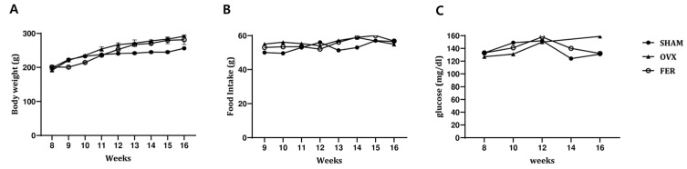
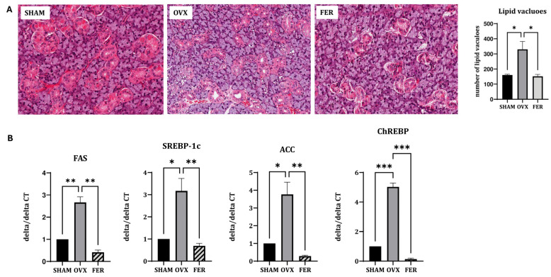
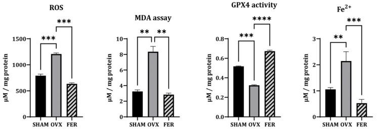
Salivary gland dysfunction is a common but underexplored complication of menopause that contributes to oral dryness, dysphagia, and increased risk of infection. Although ferroptosis, a form of regulated necrotic cell death driven by iron-dependent lipid peroxidation, has recently been implicated in postmenopausal tissue degeneration, its regulatory mechanisms in salivary glands remain unclear. In this study, we investigated the roles of mitochondrial dysfunction and mitophagy in driving ferroptosis-induced salivary gland injury in an ovariectomized (OVX) rat model of estrogen deficiency. OVX rats exhibited elevated markers of oxidative stress, lipid accumulation, and iron overload, and suppression of GPX4 activity in the salivary glands, consistent with ferroptotic activation. These changes were accompanied by impaired mitochondrial dynamics (MFN1 and OPA1), decreased expression of mitochondrial antioxidant regulators (PGC-1α, SOD, and catalase), and upregulation of mitophagy-related genes (PINK1, ULK1, Rab9, and LC3B), as well as LAMP, a lysosomal marker involved in autophagosome-lysosome fusion, while ferritinophagy (NCOA4) remained unchanged. Early administration of ferrostatin-1 effectively suppressed these pathological changes, preserving both glandular structure and function, as evidenced by the restored AQP5 and AMY2A expression. Collectively, our findings reveal that ferroptosis in estrogen-deficient salivary glands is regulated by mitochondrial instability and aberrant mitophagy, and ferrostatin-1 mitigates this cascade through multi-level mitochondrial protection. These results highlight ferrostatin-1 as a promising preventive agent against menopause-associated salivary gland dysfunction, with broader implications for organ-specific ferroptosis modulation.
AntioxidantsBiochemistry, Genetics and Molecular Biology-Physiology
CiteScore
10.60
自引率
11.40%
发文量
2123
审稿时长
16.3 days
期刊介绍:
Antioxidants (ISSN 2076-3921), provides an advanced forum for studies related to the science and technology of antioxidants. It publishes research papers, reviews and communications. Our aim is to encourage scientists to publish their experimental and theoretical results in as much detail as possible. There is no restriction on the length of the papers. The full experimental details must be provided so that the results can be reproduced. Electronic files and software regarding the full details of the calculation or experimental procedure, if unable to be published in a normal way, can be deposited as supplementary electronic material.
 求助内容:
求助内容: 应助结果提醒方式:


