Matteo Ferrante, Marianna Inglese, Ludovica Brusaferri, Nicola Toschi, Marco L Loggia
{"title":"从结构MRI图像生成合成TSPO PET图。","authors":"Matteo Ferrante, Marianna Inglese, Ludovica Brusaferri, Nicola Toschi, Marco L Loggia","doi":"10.3389/fninf.2025.1633273","DOIUrl":null,"url":null,"abstract":"<p><strong>Introduction: </strong>Neuroinflammation, a pathophysiological process involved in numerous disorders, is typically imaged using [<sup>11</sup>C]PBR28 (or TSPO) PET. However, this technique is limited by high costs and ionizing radiation, restricting its widespread clinical use. MRI, a more accessible alternative, is commonly used for structural or functional imaging, but when used using traditional approaches has limited sensitivity to specific molecular processes. This study aims to develop a deep learning model to generate TSPO PET images from structural MRI data collected in human subjects.</p><p><strong>Methods: </strong>A total of 204 scans, from participants with knee osteoarthritis (<i>n</i> = 15 scanned once, 15 scanned twice, 14 scanned three times), back pain (<i>n</i> = 40 scanned twice, 3 scanned three times), and healthy controls (<i>n</i> = 28, scanned once), underwent simultaneous 3 T MRI and [<sup>11</sup>C]PBR28 TSPO PET scans. A 3D U-Net model was trained on 80% of these PET-MRI pairs and validated using 5-fold cross-validation. The model's accuracy in reconstructed PET from MRI only was assessed using various intensity and noise metrics.</p><p><strong>Results: </strong>The model achieved a low voxel-wise mean squared error (0.0033 ± 0.0010) across all folds and a median contrast-to-noise ratio of 0.0640 ± 0.2500 when comparing true to reconstructed PET images. The synthesized PET images accurately replicated the spatial patterns observed in the original PET data. Additionally, the reconstruction accuracy was maintained even after spatial normalization.</p><p><strong>Discussion: </strong>This study demonstrates that deep learning can accurately synthesize TSPO PET images from conventional, T1-weighted MRI. This approach could enable low-cost, noninvasive neuroinflammation imaging, expanding the clinical applicability of this imaging method.</p>","PeriodicalId":12462,"journal":{"name":"Frontiers in Neuroinformatics","volume":"19 ","pages":"1633273"},"PeriodicalIF":2.5000,"publicationDate":"2025-09-08","publicationTypes":"Journal Article","fieldsOfStudy":null,"isOpenAccess":false,"openAccessPdf":"https://www.ncbi.nlm.nih.gov/pmc/articles/PMC12450709/pdf/","citationCount":"0","resultStr":"{\"title\":\"Generation of synthetic TSPO PET maps from structural MRI images.\",\"authors\":\"Matteo Ferrante, Marianna Inglese, Ludovica Brusaferri, Nicola Toschi, Marco L Loggia\",\"doi\":\"10.3389/fninf.2025.1633273\",\"DOIUrl\":null,\"url\":null,\"abstract\":\"<p><strong>Introduction: </strong>Neuroinflammation, a pathophysiological process involved in numerous disorders, is typically imaged using [<sup>11</sup>C]PBR28 (or TSPO) PET. However, this technique is limited by high costs and ionizing radiation, restricting its widespread clinical use. MRI, a more accessible alternative, is commonly used for structural or functional imaging, but when used using traditional approaches has limited sensitivity to specific molecular processes. This study aims to develop a deep learning model to generate TSPO PET images from structural MRI data collected in human subjects.</p><p><strong>Methods: </strong>A total of 204 scans, from participants with knee osteoarthritis (<i>n</i> = 15 scanned once, 15 scanned twice, 14 scanned three times), back pain (<i>n</i> = 40 scanned twice, 3 scanned three times), and healthy controls (<i>n</i> = 28, scanned once), underwent simultaneous 3 T MRI and [<sup>11</sup>C]PBR28 TSPO PET scans. A 3D U-Net model was trained on 80% of these PET-MRI pairs and validated using 5-fold cross-validation. The model's accuracy in reconstructed PET from MRI only was assessed using various intensity and noise metrics.</p><p><strong>Results: </strong>The model achieved a low voxel-wise mean squared error (0.0033 ± 0.0010) across all folds and a median contrast-to-noise ratio of 0.0640 ± 0.2500 when comparing true to reconstructed PET images. The synthesized PET images accurately replicated the spatial patterns observed in the original PET data. Additionally, the reconstruction accuracy was maintained even after spatial normalization.</p><p><strong>Discussion: </strong>This study demonstrates that deep learning can accurately synthesize TSPO PET images from conventional, T1-weighted MRI. This approach could enable low-cost, noninvasive neuroinflammation imaging, expanding the clinical applicability of this imaging method.</p>\",\"PeriodicalId\":12462,\"journal\":{\"name\":\"Frontiers in Neuroinformatics\",\"volume\":\"19 \",\"pages\":\"1633273\"},\"PeriodicalIF\":2.5000,\"publicationDate\":\"2025-09-08\",\"publicationTypes\":\"Journal Article\",\"fieldsOfStudy\":null,\"isOpenAccess\":false,\"openAccessPdf\":\"https://www.ncbi.nlm.nih.gov/pmc/articles/PMC12450709/pdf/\",\"citationCount\":\"0\",\"resultStr\":null,\"platform\":\"Semanticscholar\",\"paperid\":null,\"PeriodicalName\":\"Frontiers in Neuroinformatics\",\"FirstCategoryId\":\"3\",\"ListUrlMain\":\"https://doi.org/10.3389/fninf.2025.1633273\",\"RegionNum\":4,\"RegionCategory\":\"医学\",\"ArticlePicture\":[],\"TitleCN\":null,\"AbstractTextCN\":null,\"PMCID\":null,\"EPubDate\":\"2025/1/1 0:00:00\",\"PubModel\":\"eCollection\",\"JCR\":\"Q2\",\"JCRName\":\"MATHEMATICAL & COMPUTATIONAL BIOLOGY\",\"Score\":null,\"Total\":0}","platform":"Semanticscholar","paperid":null,"PeriodicalName":"Frontiers in Neuroinformatics","FirstCategoryId":"3","ListUrlMain":"https://doi.org/10.3389/fninf.2025.1633273","RegionNum":4,"RegionCategory":"医学","ArticlePicture":[],"TitleCN":null,"AbstractTextCN":null,"PMCID":null,"EPubDate":"2025/1/1 0:00:00","PubModel":"eCollection","JCR":"Q2","JCRName":"MATHEMATICAL & COMPUTATIONAL BIOLOGY","Score":null,"Total":0}
Generation of synthetic TSPO PET maps from structural MRI images.
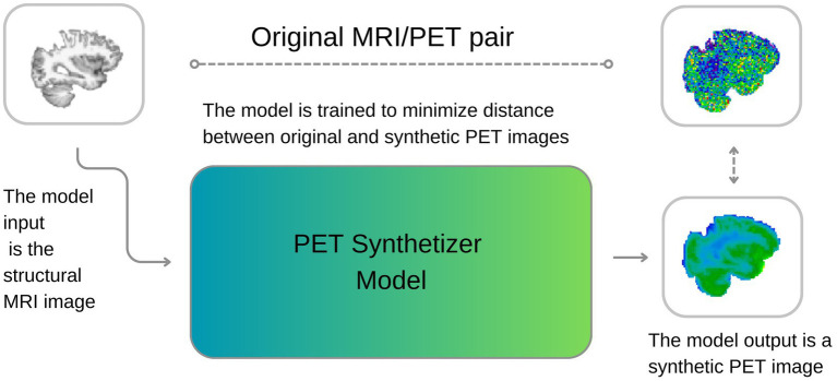
Introduction: Neuroinflammation, a pathophysiological process involved in numerous disorders, is typically imaged using [11C]PBR28 (or TSPO) PET. However, this technique is limited by high costs and ionizing radiation, restricting its widespread clinical use. MRI, a more accessible alternative, is commonly used for structural or functional imaging, but when used using traditional approaches has limited sensitivity to specific molecular processes. This study aims to develop a deep learning model to generate TSPO PET images from structural MRI data collected in human subjects.
Methods: A total of 204 scans, from participants with knee osteoarthritis (n = 15 scanned once, 15 scanned twice, 14 scanned three times), back pain (n = 40 scanned twice, 3 scanned three times), and healthy controls (n = 28, scanned once), underwent simultaneous 3 T MRI and [11C]PBR28 TSPO PET scans. A 3D U-Net model was trained on 80% of these PET-MRI pairs and validated using 5-fold cross-validation. The model's accuracy in reconstructed PET from MRI only was assessed using various intensity and noise metrics.
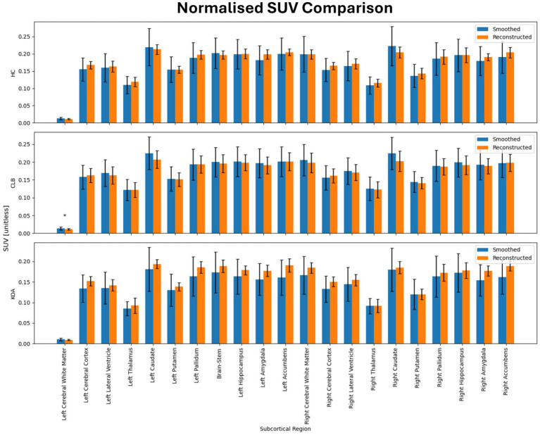
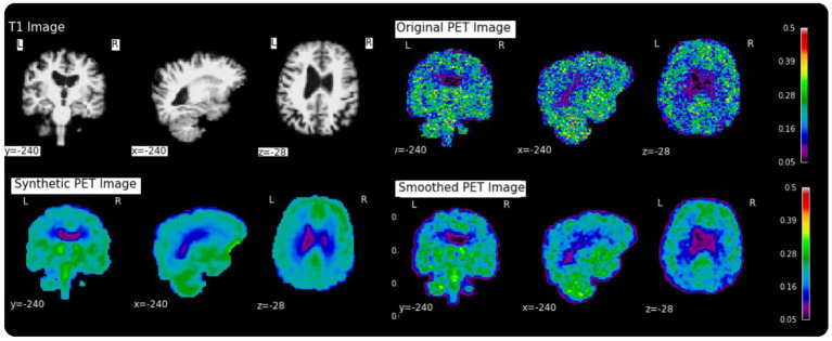
Results: The model achieved a low voxel-wise mean squared error (0.0033 ± 0.0010) across all folds and a median contrast-to-noise ratio of 0.0640 ± 0.2500 when comparing true to reconstructed PET images. The synthesized PET images accurately replicated the spatial patterns observed in the original PET data. Additionally, the reconstruction accuracy was maintained even after spatial normalization.
Discussion: This study demonstrates that deep learning can accurately synthesize TSPO PET images from conventional, T1-weighted MRI. This approach could enable low-cost, noninvasive neuroinflammation imaging, expanding the clinical applicability of this imaging method.
期刊介绍:
Frontiers in Neuroinformatics publishes rigorously peer-reviewed research on the development and implementation of numerical/computational models and analytical tools used to share, integrate and analyze experimental data and advance theories of the nervous system functions. Specialty Chief Editors Jan G. Bjaalie at the University of Oslo and Sean L. Hill at the École Polytechnique Fédérale de Lausanne are supported by an outstanding Editorial Board of international experts. This multidisciplinary open-access journal is at the forefront of disseminating and communicating scientific knowledge and impactful discoveries to researchers, academics and the public worldwide.
Neuroscience is being propelled into the information age as the volume of information explodes, demanding organization and synthesis. Novel synthesis approaches are opening up a new dimension for the exploration of the components of brain elements and systems and the vast number of variables that underlie their functions. Neural data is highly heterogeneous with complex inter-relations across multiple levels, driving the need for innovative organizing and synthesizing approaches from genes to cognition, and covering a range of species and disease states.
Frontiers in Neuroinformatics therefore welcomes submissions on existing neuroscience databases, development of data and knowledge bases for all levels of neuroscience, applications and technologies that can facilitate data sharing (interoperability, formats, terminologies, and ontologies), and novel tools for data acquisition, analyses, visualization, and dissemination of nervous system data. Our journal welcomes submissions on new tools (software and hardware) that support brain modeling, and the merging of neuroscience databases with brain models used for simulation and visualization.

 求助内容:
求助内容: 应助结果提醒方式:


