Amir F Al-Bakri, Ahmed Tahseen Muslim, Moneer K Faraj, Wamedh Esam Matti, Radana Vilimkova Kahankova, Dariusz Mikolajewski, Waldemar Karwowski, Aleksandra Kawala-Sterniuk
{"title":"在资源有限的情况下,由基于数字的符号学和VEEG支持的源定位CLARA的癫痫脑成像。","authors":"Amir F Al-Bakri, Ahmed Tahseen Muslim, Moneer K Faraj, Wamedh Esam Matti, Radana Vilimkova Kahankova, Dariusz Mikolajewski, Waldemar Karwowski, Aleksandra Kawala-Sterniuk","doi":"10.3389/fninf.2025.1661617","DOIUrl":null,"url":null,"abstract":"<p><strong>Introduction: </strong>Accurate localization of the epileptogenic zone is essential for surgical treatment of drug-resistant epilepsy. Standard presurgical evaluations rely on multimodal neuroimaging techniques, but these may be limited by availability and interpretive challenges. This study aimed to assess the concordance between zones identified by ictal semiology and a novel distributed electrical source localization technique, CLARA, and to evaluate their impact on postsurgical outcomes.</p><p><strong>Methods: </strong>This retrospective study included 16 patients with at least three recorded seizures. Ictal semiology was analyzed subjectively using video electroencephalography (VEEG) by a multidisciplinary team of neurologists, neurophysiologists, and radiologists, who determined the presumed epileptogenic zone at the lobar level. CLARA was subsequently applied to identify the computed zone based on ictal and/or interictal biomarker activities. The concordance between the presumed and computed zones was assessed qualitatively. Postsurgical outcomes were examined in relation to the extent of resection of the CLARA-defined zones.</p><p><strong>Results: </strong>Among thirteen patients with sufficient data for analysis, qualitative comparison showed 77% concordance and 23% partial concordance between the presumed and computed zones. Postsurgical follow-up revealed seizure freedom in one patient with cavernoma following complete resection of the CLARA-defined zone. In contrast, patients with incomplete resection of this region continued to experience seizures.</p><p><strong>Discussion: </strong>The findings support the potential value of CLARA as an adjunctive neuroimaging technique in the presurgical evaluation of epilepsy. By providing an additional layer of verification, CLARA may improve the accuracy of epileptogenic zone localization when used alongside established modalities such as PET, SPECT, fMRI, and MRI. Its adaptability and lower resource requirements suggest particular utility in centers with limited access to advanced medical equipment and specialized personnel. Broader implementation of CLARA could enhance presurgical decision-making and contribute to improved surgical outcomes for epilepsy patients.</p>","PeriodicalId":12462,"journal":{"name":"Frontiers in Neuroinformatics","volume":"19 ","pages":"1661617"},"PeriodicalIF":2.5000,"publicationDate":"2025-08-29","publicationTypes":"Journal Article","fieldsOfStudy":null,"isOpenAccess":false,"openAccessPdf":"https://www.ncbi.nlm.nih.gov/pmc/articles/PMC12426196/pdf/","citationCount":"0","resultStr":"{\"title\":\"Epileptic brain imaging by source localization CLARA supported by ictal-based semiology and VEEG in resource-limited settings.\",\"authors\":\"Amir F Al-Bakri, Ahmed Tahseen Muslim, Moneer K Faraj, Wamedh Esam Matti, Radana Vilimkova Kahankova, Dariusz Mikolajewski, Waldemar Karwowski, Aleksandra Kawala-Sterniuk\",\"doi\":\"10.3389/fninf.2025.1661617\",\"DOIUrl\":null,\"url\":null,\"abstract\":\"<p><strong>Introduction: </strong>Accurate localization of the epileptogenic zone is essential for surgical treatment of drug-resistant epilepsy. Standard presurgical evaluations rely on multimodal neuroimaging techniques, but these may be limited by availability and interpretive challenges. This study aimed to assess the concordance between zones identified by ictal semiology and a novel distributed electrical source localization technique, CLARA, and to evaluate their impact on postsurgical outcomes.</p><p><strong>Methods: </strong>This retrospective study included 16 patients with at least three recorded seizures. Ictal semiology was analyzed subjectively using video electroencephalography (VEEG) by a multidisciplinary team of neurologists, neurophysiologists, and radiologists, who determined the presumed epileptogenic zone at the lobar level. CLARA was subsequently applied to identify the computed zone based on ictal and/or interictal biomarker activities. The concordance between the presumed and computed zones was assessed qualitatively. Postsurgical outcomes were examined in relation to the extent of resection of the CLARA-defined zones.</p><p><strong>Results: </strong>Among thirteen patients with sufficient data for analysis, qualitative comparison showed 77% concordance and 23% partial concordance between the presumed and computed zones. Postsurgical follow-up revealed seizure freedom in one patient with cavernoma following complete resection of the CLARA-defined zone. In contrast, patients with incomplete resection of this region continued to experience seizures.</p><p><strong>Discussion: </strong>The findings support the potential value of CLARA as an adjunctive neuroimaging technique in the presurgical evaluation of epilepsy. By providing an additional layer of verification, CLARA may improve the accuracy of epileptogenic zone localization when used alongside established modalities such as PET, SPECT, fMRI, and MRI. Its adaptability and lower resource requirements suggest particular utility in centers with limited access to advanced medical equipment and specialized personnel. Broader implementation of CLARA could enhance presurgical decision-making and contribute to improved surgical outcomes for epilepsy patients.</p>\",\"PeriodicalId\":12462,\"journal\":{\"name\":\"Frontiers in Neuroinformatics\",\"volume\":\"19 \",\"pages\":\"1661617\"},\"PeriodicalIF\":2.5000,\"publicationDate\":\"2025-08-29\",\"publicationTypes\":\"Journal Article\",\"fieldsOfStudy\":null,\"isOpenAccess\":false,\"openAccessPdf\":\"https://www.ncbi.nlm.nih.gov/pmc/articles/PMC12426196/pdf/\",\"citationCount\":\"0\",\"resultStr\":null,\"platform\":\"Semanticscholar\",\"paperid\":null,\"PeriodicalName\":\"Frontiers in Neuroinformatics\",\"FirstCategoryId\":\"3\",\"ListUrlMain\":\"https://doi.org/10.3389/fninf.2025.1661617\",\"RegionNum\":4,\"RegionCategory\":\"医学\",\"ArticlePicture\":[],\"TitleCN\":null,\"AbstractTextCN\":null,\"PMCID\":null,\"EPubDate\":\"2025/1/1 0:00:00\",\"PubModel\":\"eCollection\",\"JCR\":\"Q2\",\"JCRName\":\"MATHEMATICAL & COMPUTATIONAL BIOLOGY\",\"Score\":null,\"Total\":0}","platform":"Semanticscholar","paperid":null,"PeriodicalName":"Frontiers in Neuroinformatics","FirstCategoryId":"3","ListUrlMain":"https://doi.org/10.3389/fninf.2025.1661617","RegionNum":4,"RegionCategory":"医学","ArticlePicture":[],"TitleCN":null,"AbstractTextCN":null,"PMCID":null,"EPubDate":"2025/1/1 0:00:00","PubModel":"eCollection","JCR":"Q2","JCRName":"MATHEMATICAL & COMPUTATIONAL BIOLOGY","Score":null,"Total":0}
Epileptic brain imaging by source localization CLARA supported by ictal-based semiology and VEEG in resource-limited settings.
Introduction: Accurate localization of the epileptogenic zone is essential for surgical treatment of drug-resistant epilepsy. Standard presurgical evaluations rely on multimodal neuroimaging techniques, but these may be limited by availability and interpretive challenges. This study aimed to assess the concordance between zones identified by ictal semiology and a novel distributed electrical source localization technique, CLARA, and to evaluate their impact on postsurgical outcomes.
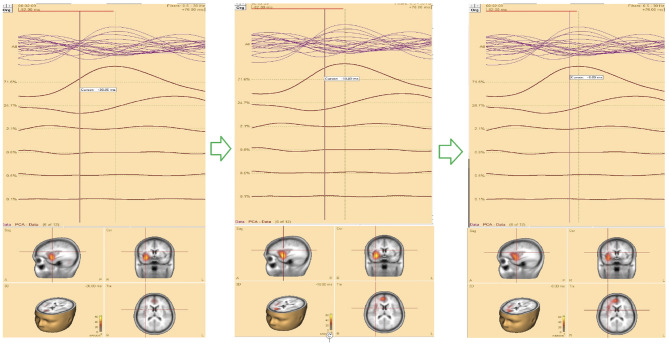
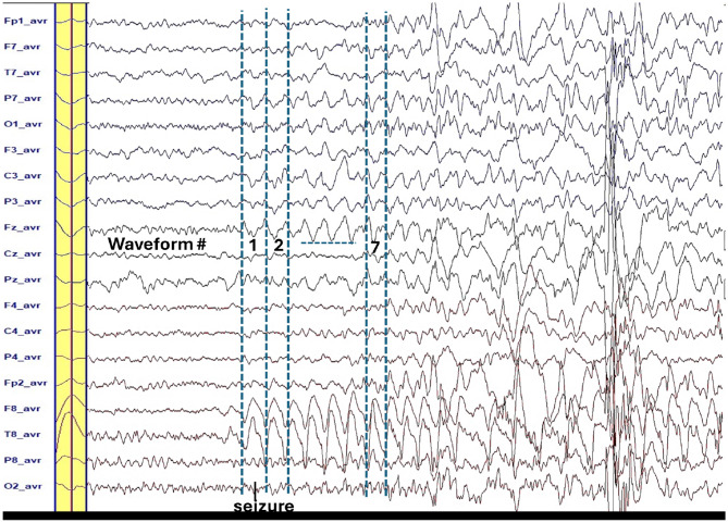
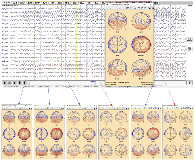
Methods: This retrospective study included 16 patients with at least three recorded seizures. Ictal semiology was analyzed subjectively using video electroencephalography (VEEG) by a multidisciplinary team of neurologists, neurophysiologists, and radiologists, who determined the presumed epileptogenic zone at the lobar level. CLARA was subsequently applied to identify the computed zone based on ictal and/or interictal biomarker activities. The concordance between the presumed and computed zones was assessed qualitatively. Postsurgical outcomes were examined in relation to the extent of resection of the CLARA-defined zones.
Results: Among thirteen patients with sufficient data for analysis, qualitative comparison showed 77% concordance and 23% partial concordance between the presumed and computed zones. Postsurgical follow-up revealed seizure freedom in one patient with cavernoma following complete resection of the CLARA-defined zone. In contrast, patients with incomplete resection of this region continued to experience seizures.
Discussion: The findings support the potential value of CLARA as an adjunctive neuroimaging technique in the presurgical evaluation of epilepsy. By providing an additional layer of verification, CLARA may improve the accuracy of epileptogenic zone localization when used alongside established modalities such as PET, SPECT, fMRI, and MRI. Its adaptability and lower resource requirements suggest particular utility in centers with limited access to advanced medical equipment and specialized personnel. Broader implementation of CLARA could enhance presurgical decision-making and contribute to improved surgical outcomes for epilepsy patients.
期刊介绍:
Frontiers in Neuroinformatics publishes rigorously peer-reviewed research on the development and implementation of numerical/computational models and analytical tools used to share, integrate and analyze experimental data and advance theories of the nervous system functions. Specialty Chief Editors Jan G. Bjaalie at the University of Oslo and Sean L. Hill at the École Polytechnique Fédérale de Lausanne are supported by an outstanding Editorial Board of international experts. This multidisciplinary open-access journal is at the forefront of disseminating and communicating scientific knowledge and impactful discoveries to researchers, academics and the public worldwide.
Neuroscience is being propelled into the information age as the volume of information explodes, demanding organization and synthesis. Novel synthesis approaches are opening up a new dimension for the exploration of the components of brain elements and systems and the vast number of variables that underlie their functions. Neural data is highly heterogeneous with complex inter-relations across multiple levels, driving the need for innovative organizing and synthesizing approaches from genes to cognition, and covering a range of species and disease states.
Frontiers in Neuroinformatics therefore welcomes submissions on existing neuroscience databases, development of data and knowledge bases for all levels of neuroscience, applications and technologies that can facilitate data sharing (interoperability, formats, terminologies, and ontologies), and novel tools for data acquisition, analyses, visualization, and dissemination of nervous system data. Our journal welcomes submissions on new tools (software and hardware) that support brain modeling, and the merging of neuroscience databases with brain models used for simulation and visualization.

 求助内容:
求助内容: 应助结果提醒方式:


