{"title":"光暗条件下皮层视觉层次的时间敏感性。","authors":"Deena Elul, Ayelet McKyton, Netta Levin","doi":"10.1167/iovs.66.12.12","DOIUrl":null,"url":null,"abstract":"<p><strong>Purpose: </strong>Behavioral and electrophysiological studies have shown that vision is slower under scotopic conditions (dark, activating only rods) than photopic conditions (light, activating only cones). However, slower scotopic processing cannot be solely explained by findings that rod signals are slower than cone signals, and it is unknown whether temporal processing differences persist in cortex. Flickering stimuli have previously been used in functional MRI (fMRI) studies to probe photopic cortical temporal sensitivity. This fMRI study investigates flicker sensitivity under photopic and scotopic conditions across the cortical visual hierarchy.</p><p><strong>Methods: </strong>Fourteen participants viewed a stimulus flickering at six frequencies (2-10 Hz) under photopic and scotopic conditions during fMRI scanning. Retinotopic and high-level visual areas were delineated for each subject with population receptive field modeling (using a drifting bar) and a functional localizer (using images of objects).</p><p><strong>Results: </strong>In most areas, higher mean activation was observed under photopic than under scotopic conditions. However, peak activation was higher only in V1 and ventral retinotopic areas. The pattern of change over frequencies differed between lighting conditions in retinotopic areas, but not in most high-level areas. Under scotopic conditions, the largest BOLD response was observed at low frequencies throughout visual cortex. Under photopic conditions, BOLD responses appeared largely unchanging across frequencies, with a trend towards preferring higher frequencies in V1.</p><p><strong>Conclusions: </strong>Selectivity for lower frequencies under scotopic conditions was observed throughout visual cortex, in contrast to limited selectivity under photopic conditions. This low-frequency preference could allow more time for extracting information from sparse scotopic stimuli.</p>","PeriodicalId":14620,"journal":{"name":"Investigative ophthalmology & visual science","volume":"66 12","pages":"12"},"PeriodicalIF":4.7000,"publicationDate":"2025-09-02","publicationTypes":"Journal Article","fieldsOfStudy":null,"isOpenAccess":false,"openAccessPdf":"https://www.ncbi.nlm.nih.gov/pmc/articles/PMC12416517/pdf/","citationCount":"0","resultStr":"{\"title\":\"Temporal Sensitivity Under Photopic and Scotopic Conditions Across the Cortical Visual Hierarchy.\",\"authors\":\"Deena Elul, Ayelet McKyton, Netta Levin\",\"doi\":\"10.1167/iovs.66.12.12\",\"DOIUrl\":null,\"url\":null,\"abstract\":\"<p><strong>Purpose: </strong>Behavioral and electrophysiological studies have shown that vision is slower under scotopic conditions (dark, activating only rods) than photopic conditions (light, activating only cones). However, slower scotopic processing cannot be solely explained by findings that rod signals are slower than cone signals, and it is unknown whether temporal processing differences persist in cortex. Flickering stimuli have previously been used in functional MRI (fMRI) studies to probe photopic cortical temporal sensitivity. This fMRI study investigates flicker sensitivity under photopic and scotopic conditions across the cortical visual hierarchy.</p><p><strong>Methods: </strong>Fourteen participants viewed a stimulus flickering at six frequencies (2-10 Hz) under photopic and scotopic conditions during fMRI scanning. Retinotopic and high-level visual areas were delineated for each subject with population receptive field modeling (using a drifting bar) and a functional localizer (using images of objects).</p><p><strong>Results: </strong>In most areas, higher mean activation was observed under photopic than under scotopic conditions. However, peak activation was higher only in V1 and ventral retinotopic areas. The pattern of change over frequencies differed between lighting conditions in retinotopic areas, but not in most high-level areas. Under scotopic conditions, the largest BOLD response was observed at low frequencies throughout visual cortex. Under photopic conditions, BOLD responses appeared largely unchanging across frequencies, with a trend towards preferring higher frequencies in V1.</p><p><strong>Conclusions: </strong>Selectivity for lower frequencies under scotopic conditions was observed throughout visual cortex, in contrast to limited selectivity under photopic conditions. This low-frequency preference could allow more time for extracting information from sparse scotopic stimuli.</p>\",\"PeriodicalId\":14620,\"journal\":{\"name\":\"Investigative ophthalmology & visual science\",\"volume\":\"66 12\",\"pages\":\"12\"},\"PeriodicalIF\":4.7000,\"publicationDate\":\"2025-09-02\",\"publicationTypes\":\"Journal Article\",\"fieldsOfStudy\":null,\"isOpenAccess\":false,\"openAccessPdf\":\"https://www.ncbi.nlm.nih.gov/pmc/articles/PMC12416517/pdf/\",\"citationCount\":\"0\",\"resultStr\":null,\"platform\":\"Semanticscholar\",\"paperid\":null,\"PeriodicalName\":\"Investigative ophthalmology & visual science\",\"FirstCategoryId\":\"3\",\"ListUrlMain\":\"https://doi.org/10.1167/iovs.66.12.12\",\"RegionNum\":2,\"RegionCategory\":\"医学\",\"ArticlePicture\":[],\"TitleCN\":null,\"AbstractTextCN\":null,\"PMCID\":null,\"EPubDate\":\"\",\"PubModel\":\"\",\"JCR\":\"Q1\",\"JCRName\":\"OPHTHALMOLOGY\",\"Score\":null,\"Total\":0}","platform":"Semanticscholar","paperid":null,"PeriodicalName":"Investigative ophthalmology & visual science","FirstCategoryId":"3","ListUrlMain":"https://doi.org/10.1167/iovs.66.12.12","RegionNum":2,"RegionCategory":"医学","ArticlePicture":[],"TitleCN":null,"AbstractTextCN":null,"PMCID":null,"EPubDate":"","PubModel":"","JCR":"Q1","JCRName":"OPHTHALMOLOGY","Score":null,"Total":0}
Temporal Sensitivity Under Photopic and Scotopic Conditions Across the Cortical Visual Hierarchy.
Purpose: Behavioral and electrophysiological studies have shown that vision is slower under scotopic conditions (dark, activating only rods) than photopic conditions (light, activating only cones). However, slower scotopic processing cannot be solely explained by findings that rod signals are slower than cone signals, and it is unknown whether temporal processing differences persist in cortex. Flickering stimuli have previously been used in functional MRI (fMRI) studies to probe photopic cortical temporal sensitivity. This fMRI study investigates flicker sensitivity under photopic and scotopic conditions across the cortical visual hierarchy.
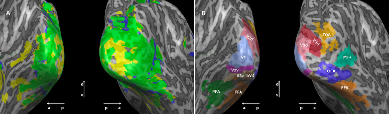
Methods: Fourteen participants viewed a stimulus flickering at six frequencies (2-10 Hz) under photopic and scotopic conditions during fMRI scanning. Retinotopic and high-level visual areas were delineated for each subject with population receptive field modeling (using a drifting bar) and a functional localizer (using images of objects).
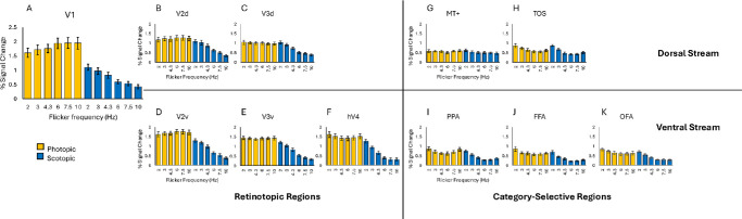
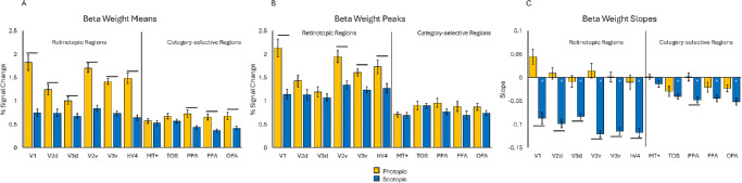
Results: In most areas, higher mean activation was observed under photopic than under scotopic conditions. However, peak activation was higher only in V1 and ventral retinotopic areas. The pattern of change over frequencies differed between lighting conditions in retinotopic areas, but not in most high-level areas. Under scotopic conditions, the largest BOLD response was observed at low frequencies throughout visual cortex. Under photopic conditions, BOLD responses appeared largely unchanging across frequencies, with a trend towards preferring higher frequencies in V1.
Conclusions: Selectivity for lower frequencies under scotopic conditions was observed throughout visual cortex, in contrast to limited selectivity under photopic conditions. This low-frequency preference could allow more time for extracting information from sparse scotopic stimuli.
期刊介绍:
Investigative Ophthalmology & Visual Science (IOVS), published as ready online, is a peer-reviewed academic journal of the Association for Research in Vision and Ophthalmology (ARVO). IOVS features original research, mostly pertaining to clinical and laboratory ophthalmology and vision research in general.

 求助内容:
求助内容: 应助结果提醒方式:


