Clancy Cerejo, Elias Mandler, Federico Carbone, Gabriel Bsteh, Barbara Teuchner, Katarína Schwarzová, Marina Peball, Atbin Djamshidian, Klaus Seppi, Beatrice Heim
{"title":"光学相干断层扫描-早期亨廷顿氏病的可能生物标志物。","authors":"Clancy Cerejo, Elias Mandler, Federico Carbone, Gabriel Bsteh, Barbara Teuchner, Katarína Schwarzová, Marina Peball, Atbin Djamshidian, Klaus Seppi, Beatrice Heim","doi":"10.1186/s42466-025-00421-z","DOIUrl":null,"url":null,"abstract":"<p><strong>Objective: </strong>To assess the role of spectral domain Optical Coherence Tomography (OCT) as a biomarker in Huntington's disease (HD).</p><p><strong>Methods: </strong>This cross-sectional study compared spectral domain OCT data, cognitive function, and olfactory function in HD patients and healthy controls (HC). HD patients were classified into Stage1 and Stage2 based on motor symptoms and functional capacity.</p><p><strong>Results: </strong>We recruited a total of 68 participants including 39HD patients (22 stage1, 17 stage2) and 29 age-matched HC. There were no significant differences in age and gender between the groups. Stage2 HD patients showed worse motor function (UHDRS-TMS 28.44 ± 18.13 vs. 13.74 ± 8.78, p = 0.002), functional capacity (UHDRS-TFC 8.13 ± 2.03 vs. 12.44 ± 0.99, p < 0.001), and lower scores on MMSE (27.36 ± 1.64 vs. 28.73 ± 1.74, p = 0.005 vs. 29.45 ± 0.91, p < 0.001) compared to stage1 HD patients and HC, respectively. Both stage1 and stage2 HD groups displayed significantly reduced macular retinal nerve fibre layer thickness (mRNFL) (33.45 ± 4.70, 31.90 ± 3.47 vs. 38.45 ± 5.00; p < 0.001) and ganglion cell-inner plexiform layer thickness (GCIPL) (71.63 ± 6.38, p = 0.007; 60.42 ± 4.67, p < 0.001 vs. 77.03 ± 8.40) as compared to HC. The retinal OCT parameters mRNFL and GCIPL correlated moderately with PIN<sub>HD</sub> (r=-0.424, r=-0.513; p < 0.001), CAP (r=-0.425, r=-0.482; p < 0.001) and olfactory dysfunction for both smell identification (r = 0.446, r = 0.500; p < 0.001) and smell discrimination (r = 0.563, r = 0.467; p < 0.001).</p><p><strong>Conclusions: </strong>HD patients exhibit significantly thinner retinal ganglion cell inner plexiform layer and macular retinal nerve fibre layer compared to HC, even in the early phase of the disease. These findings suggest that OCT may serve as a valuable biomarker to monitor neurodegeneration at an early disease stage.</p>","PeriodicalId":94156,"journal":{"name":"Neurological research and practice","volume":"7 1","pages":"61"},"PeriodicalIF":3.2000,"publicationDate":"2025-08-28","publicationTypes":"Journal Article","fieldsOfStudy":null,"isOpenAccess":false,"openAccessPdf":"https://www.ncbi.nlm.nih.gov/pmc/articles/PMC12395900/pdf/","citationCount":"0","resultStr":"{\"title\":\"Optical coherence tomography - A possible biomarker in early huntington's disease.\",\"authors\":\"Clancy Cerejo, Elias Mandler, Federico Carbone, Gabriel Bsteh, Barbara Teuchner, Katarína Schwarzová, Marina Peball, Atbin Djamshidian, Klaus Seppi, Beatrice Heim\",\"doi\":\"10.1186/s42466-025-00421-z\",\"DOIUrl\":null,\"url\":null,\"abstract\":\"<p><strong>Objective: </strong>To assess the role of spectral domain Optical Coherence Tomography (OCT) as a biomarker in Huntington's disease (HD).</p><p><strong>Methods: </strong>This cross-sectional study compared spectral domain OCT data, cognitive function, and olfactory function in HD patients and healthy controls (HC). HD patients were classified into Stage1 and Stage2 based on motor symptoms and functional capacity.</p><p><strong>Results: </strong>We recruited a total of 68 participants including 39HD patients (22 stage1, 17 stage2) and 29 age-matched HC. There were no significant differences in age and gender between the groups. Stage2 HD patients showed worse motor function (UHDRS-TMS 28.44 ± 18.13 vs. 13.74 ± 8.78, p = 0.002), functional capacity (UHDRS-TFC 8.13 ± 2.03 vs. 12.44 ± 0.99, p < 0.001), and lower scores on MMSE (27.36 ± 1.64 vs. 28.73 ± 1.74, p = 0.005 vs. 29.45 ± 0.91, p < 0.001) compared to stage1 HD patients and HC, respectively. Both stage1 and stage2 HD groups displayed significantly reduced macular retinal nerve fibre layer thickness (mRNFL) (33.45 ± 4.70, 31.90 ± 3.47 vs. 38.45 ± 5.00; p < 0.001) and ganglion cell-inner plexiform layer thickness (GCIPL) (71.63 ± 6.38, p = 0.007; 60.42 ± 4.67, p < 0.001 vs. 77.03 ± 8.40) as compared to HC. The retinal OCT parameters mRNFL and GCIPL correlated moderately with PIN<sub>HD</sub> (r=-0.424, r=-0.513; p < 0.001), CAP (r=-0.425, r=-0.482; p < 0.001) and olfactory dysfunction for both smell identification (r = 0.446, r = 0.500; p < 0.001) and smell discrimination (r = 0.563, r = 0.467; p < 0.001).</p><p><strong>Conclusions: </strong>HD patients exhibit significantly thinner retinal ganglion cell inner plexiform layer and macular retinal nerve fibre layer compared to HC, even in the early phase of the disease. These findings suggest that OCT may serve as a valuable biomarker to monitor neurodegeneration at an early disease stage.</p>\",\"PeriodicalId\":94156,\"journal\":{\"name\":\"Neurological research and practice\",\"volume\":\"7 1\",\"pages\":\"61\"},\"PeriodicalIF\":3.2000,\"publicationDate\":\"2025-08-28\",\"publicationTypes\":\"Journal Article\",\"fieldsOfStudy\":null,\"isOpenAccess\":false,\"openAccessPdf\":\"https://www.ncbi.nlm.nih.gov/pmc/articles/PMC12395900/pdf/\",\"citationCount\":\"0\",\"resultStr\":null,\"platform\":\"Semanticscholar\",\"paperid\":null,\"PeriodicalName\":\"Neurological research and practice\",\"FirstCategoryId\":\"1085\",\"ListUrlMain\":\"https://doi.org/10.1186/s42466-025-00421-z\",\"RegionNum\":0,\"RegionCategory\":null,\"ArticlePicture\":[],\"TitleCN\":null,\"AbstractTextCN\":null,\"PMCID\":null,\"EPubDate\":\"\",\"PubModel\":\"\",\"JCR\":\"Q2\",\"JCRName\":\"Medicine\",\"Score\":null,\"Total\":0}","platform":"Semanticscholar","paperid":null,"PeriodicalName":"Neurological research and practice","FirstCategoryId":"1085","ListUrlMain":"https://doi.org/10.1186/s42466-025-00421-z","RegionNum":0,"RegionCategory":null,"ArticlePicture":[],"TitleCN":null,"AbstractTextCN":null,"PMCID":null,"EPubDate":"","PubModel":"","JCR":"Q2","JCRName":"Medicine","Score":null,"Total":0}
引用次数: 0
摘要
目的:评价光谱域光学相干断层扫描(OCT)作为亨廷顿舞蹈病(HD)生物标志物的作用。方法:本横断面研究比较了HD患者和健康对照(HC)的光谱域OCT数据、认知功能和嗅觉功能。根据运动症状和功能能力将HD患者分为1期和2期。结果:我们共招募了68名参与者,包括39HD患者(22例1期,17例2期)和29例年龄匹配的HC。两组之间的年龄和性别没有显著差异。2期HD患者运动功能差(UHDRS-TMS 28.44±18.13 vs. 13.74±8.78,p = 0.002),功能容量差(UHDRS-TFC 8.13±2.03 vs. 12.44±0.99,p HD) (r=-0.424, r=-0.513; p)结论:HD患者即使在疾病早期,视网膜神经节细胞内丛状层和黄斑视网膜神经纤维层也明显较HC薄。这些发现表明OCT可以作为一种有价值的生物标志物,在疾病早期监测神经退行性变。
Optical coherence tomography - A possible biomarker in early huntington's disease.
Objective: To assess the role of spectral domain Optical Coherence Tomography (OCT) as a biomarker in Huntington's disease (HD).
Methods: This cross-sectional study compared spectral domain OCT data, cognitive function, and olfactory function in HD patients and healthy controls (HC). HD patients were classified into Stage1 and Stage2 based on motor symptoms and functional capacity.
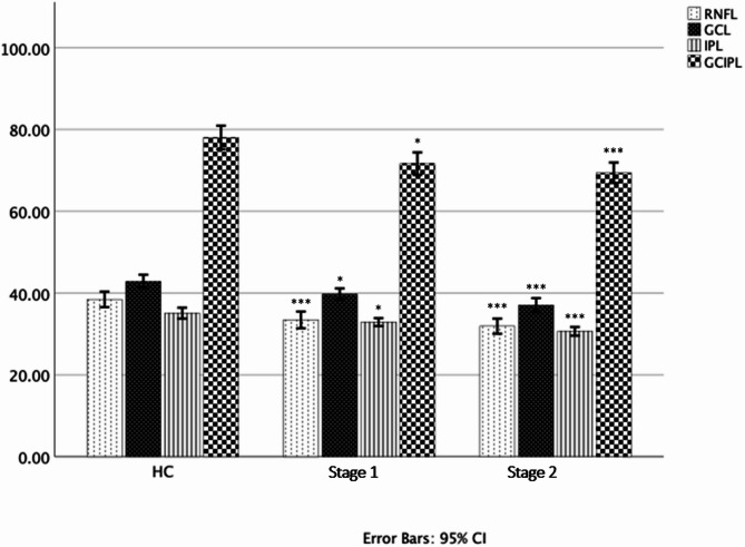
Results: We recruited a total of 68 participants including 39HD patients (22 stage1, 17 stage2) and 29 age-matched HC. There were no significant differences in age and gender between the groups. Stage2 HD patients showed worse motor function (UHDRS-TMS 28.44 ± 18.13 vs. 13.74 ± 8.78, p = 0.002), functional capacity (UHDRS-TFC 8.13 ± 2.03 vs. 12.44 ± 0.99, p < 0.001), and lower scores on MMSE (27.36 ± 1.64 vs. 28.73 ± 1.74, p = 0.005 vs. 29.45 ± 0.91, p < 0.001) compared to stage1 HD patients and HC, respectively. Both stage1 and stage2 HD groups displayed significantly reduced macular retinal nerve fibre layer thickness (mRNFL) (33.45 ± 4.70, 31.90 ± 3.47 vs. 38.45 ± 5.00; p < 0.001) and ganglion cell-inner plexiform layer thickness (GCIPL) (71.63 ± 6.38, p = 0.007; 60.42 ± 4.67, p < 0.001 vs. 77.03 ± 8.40) as compared to HC. The retinal OCT parameters mRNFL and GCIPL correlated moderately with PINHD (r=-0.424, r=-0.513; p < 0.001), CAP (r=-0.425, r=-0.482; p < 0.001) and olfactory dysfunction for both smell identification (r = 0.446, r = 0.500; p < 0.001) and smell discrimination (r = 0.563, r = 0.467; p < 0.001).
Conclusions: HD patients exhibit significantly thinner retinal ganglion cell inner plexiform layer and macular retinal nerve fibre layer compared to HC, even in the early phase of the disease. These findings suggest that OCT may serve as a valuable biomarker to monitor neurodegeneration at an early disease stage.

 求助内容:
求助内容: 应助结果提醒方式:


