{"title":"利用生理模型预测青光眼的结构-功能关系。","authors":"Chris Bradley, Jithin Yohannan","doi":"10.1167/iovs.66.11.75","DOIUrl":null,"url":null,"abstract":"<p><strong>Purpose: </strong>Determine how well a physiological model-the retina-V1 (RV1) model of target detection-predicts the structure-function relationship in glaucoma.</p><p><strong>Methods: </strong>Unlike curve-fitting models, the RV1 model includes a map of retinal ganglion cell (RGC) receptive fields across the visual field (VF), enabling simulation of different patterns of RGC loss. Predicted mean sensitivity for different patterns of simulated RGC loss and predictions of different curve-fitting models were compared to 12,917 paired SITA-Standard 24-2 VFs and optical coherence tomography measurements of average retinal nerve fiber layer thickness from 4432 eyes of 2418 patients with glaucoma between 1997 and 2023. Except for one free parameter, all RV1 model parameters were fixed from a previous study that fit the model to an unrelated data set of contrast sensitivities for 43 localized achromatic stimuli.</p><p><strong>Results: </strong>Different structure-function relationships were predicted by the RV1 model for different patterns of RGC loss. Random RGC loss resulted in a mean absolute error of 2.99 dB, which was marginally but significantly smaller than 3.01 dB for ninth-degree polynomial regression-the highest degree polynomial whose coefficients could be estimated. Other patterns of simulated RGC loss, including periphery-to-fovea loss typical in glaucoma, accounted for much of the observed variance in the structure-function data. Unlike curve-fitting models, the RV1 model correctly predicted higher variance at lower dB/micron levels.</p><p><strong>Conclusions: </strong>A physiological model can account for much of the observed variance in structure-function data for glaucoma by simulating different patterns of RGC loss-this is currently not possible with curve-fitting models.</p>","PeriodicalId":14620,"journal":{"name":"Investigative ophthalmology & visual science","volume":"66 11","pages":"75"},"PeriodicalIF":4.7000,"publicationDate":"2025-08-01","publicationTypes":"Journal Article","fieldsOfStudy":null,"isOpenAccess":false,"openAccessPdf":"https://www.ncbi.nlm.nih.gov/pmc/articles/PMC12400969/pdf/","citationCount":"0","resultStr":"{\"title\":\"Predicting the Structure-Function Relationship in Glaucoma Using a Physiological Model.\",\"authors\":\"Chris Bradley, Jithin Yohannan\",\"doi\":\"10.1167/iovs.66.11.75\",\"DOIUrl\":null,\"url\":null,\"abstract\":\"<p><strong>Purpose: </strong>Determine how well a physiological model-the retina-V1 (RV1) model of target detection-predicts the structure-function relationship in glaucoma.</p><p><strong>Methods: </strong>Unlike curve-fitting models, the RV1 model includes a map of retinal ganglion cell (RGC) receptive fields across the visual field (VF), enabling simulation of different patterns of RGC loss. Predicted mean sensitivity for different patterns of simulated RGC loss and predictions of different curve-fitting models were compared to 12,917 paired SITA-Standard 24-2 VFs and optical coherence tomography measurements of average retinal nerve fiber layer thickness from 4432 eyes of 2418 patients with glaucoma between 1997 and 2023. Except for one free parameter, all RV1 model parameters were fixed from a previous study that fit the model to an unrelated data set of contrast sensitivities for 43 localized achromatic stimuli.</p><p><strong>Results: </strong>Different structure-function relationships were predicted by the RV1 model for different patterns of RGC loss. Random RGC loss resulted in a mean absolute error of 2.99 dB, which was marginally but significantly smaller than 3.01 dB for ninth-degree polynomial regression-the highest degree polynomial whose coefficients could be estimated. Other patterns of simulated RGC loss, including periphery-to-fovea loss typical in glaucoma, accounted for much of the observed variance in the structure-function data. Unlike curve-fitting models, the RV1 model correctly predicted higher variance at lower dB/micron levels.</p><p><strong>Conclusions: </strong>A physiological model can account for much of the observed variance in structure-function data for glaucoma by simulating different patterns of RGC loss-this is currently not possible with curve-fitting models.</p>\",\"PeriodicalId\":14620,\"journal\":{\"name\":\"Investigative ophthalmology & visual science\",\"volume\":\"66 11\",\"pages\":\"75\"},\"PeriodicalIF\":4.7000,\"publicationDate\":\"2025-08-01\",\"publicationTypes\":\"Journal Article\",\"fieldsOfStudy\":null,\"isOpenAccess\":false,\"openAccessPdf\":\"https://www.ncbi.nlm.nih.gov/pmc/articles/PMC12400969/pdf/\",\"citationCount\":\"0\",\"resultStr\":null,\"platform\":\"Semanticscholar\",\"paperid\":null,\"PeriodicalName\":\"Investigative ophthalmology & visual science\",\"FirstCategoryId\":\"3\",\"ListUrlMain\":\"https://doi.org/10.1167/iovs.66.11.75\",\"RegionNum\":2,\"RegionCategory\":\"医学\",\"ArticlePicture\":[],\"TitleCN\":null,\"AbstractTextCN\":null,\"PMCID\":null,\"EPubDate\":\"\",\"PubModel\":\"\",\"JCR\":\"Q1\",\"JCRName\":\"OPHTHALMOLOGY\",\"Score\":null,\"Total\":0}","platform":"Semanticscholar","paperid":null,"PeriodicalName":"Investigative ophthalmology & visual science","FirstCategoryId":"3","ListUrlMain":"https://doi.org/10.1167/iovs.66.11.75","RegionNum":2,"RegionCategory":"医学","ArticlePicture":[],"TitleCN":null,"AbstractTextCN":null,"PMCID":null,"EPubDate":"","PubModel":"","JCR":"Q1","JCRName":"OPHTHALMOLOGY","Score":null,"Total":0}
Predicting the Structure-Function Relationship in Glaucoma Using a Physiological Model.
Purpose: Determine how well a physiological model-the retina-V1 (RV1) model of target detection-predicts the structure-function relationship in glaucoma.
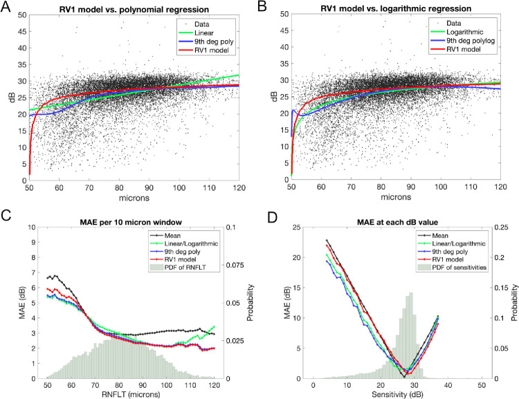
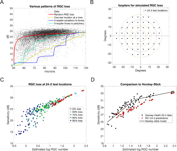
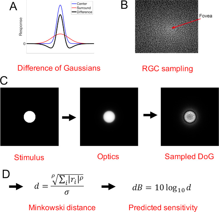
Methods: Unlike curve-fitting models, the RV1 model includes a map of retinal ganglion cell (RGC) receptive fields across the visual field (VF), enabling simulation of different patterns of RGC loss. Predicted mean sensitivity for different patterns of simulated RGC loss and predictions of different curve-fitting models were compared to 12,917 paired SITA-Standard 24-2 VFs and optical coherence tomography measurements of average retinal nerve fiber layer thickness from 4432 eyes of 2418 patients with glaucoma between 1997 and 2023. Except for one free parameter, all RV1 model parameters were fixed from a previous study that fit the model to an unrelated data set of contrast sensitivities for 43 localized achromatic stimuli.
Results: Different structure-function relationships were predicted by the RV1 model for different patterns of RGC loss. Random RGC loss resulted in a mean absolute error of 2.99 dB, which was marginally but significantly smaller than 3.01 dB for ninth-degree polynomial regression-the highest degree polynomial whose coefficients could be estimated. Other patterns of simulated RGC loss, including periphery-to-fovea loss typical in glaucoma, accounted for much of the observed variance in the structure-function data. Unlike curve-fitting models, the RV1 model correctly predicted higher variance at lower dB/micron levels.
Conclusions: A physiological model can account for much of the observed variance in structure-function data for glaucoma by simulating different patterns of RGC loss-this is currently not possible with curve-fitting models.
期刊介绍:
Investigative Ophthalmology & Visual Science (IOVS), published as ready online, is a peer-reviewed academic journal of the Association for Research in Vision and Ophthalmology (ARVO). IOVS features original research, mostly pertaining to clinical and laboratory ophthalmology and vision research in general.

 求助内容:
求助内容: 应助结果提醒方式:


