{"title":"匹配光学相干断层扫描血管造影指标对糖尿病视网膜病变严重程度检测进展的重要性。","authors":"Shinji Kakihara, Kallista Zhuang, Mohamed AbdelSalam, Taffeta Chingning Yamaguchi, Amani A Fawzi","doi":"10.1167/iovs.66.11.49","DOIUrl":null,"url":null,"abstract":"<p><strong>Purpose: </strong>The purpose of this study was to determine which macular optical coherence tomography angiography (OCTA) metric best captures early progression of capillary non-perfusion across diabetic retinopathy (DR) severities.</p><p><strong>Methods: </strong>In this prospective, 1-year observational study, 208 patients with diabetes (320 eyes) underwent 3 × 3 mm macular OCTA at baseline, 6 months, and 12 months. Registered, averaged images yielded geometric perfusion deficits (GPDs), vessel density (VD), vessel length density (VLD) in the superficial and deep capillary plexuses (SCP and DCP), and foveal avascular zone area. Eyes were graded as non-referable or referable. Linear mixed models adjusted for age, sex, diabetes duration, hemoglobin A1c, and hypertension were conducted. Post hoc Dunnett's tests compared follow-ups with baseline within each severity group.</p><p><strong>Results: </strong>A total of 709 eye visits were analyzed by OCTA. Across the cohort, referable DR, longer diabetes duration, and hypertension were independently associated with higher GPD values. In referable eyes, GPD-DCP increased at 6 months (P = 0.036) and 1 year (P = 0.016), whereas no other OCTA metric changed significantly at 6 months. In non-referable eyes, the only significant change was a decrease in VD-SCP at 1 year (P = 0.004).</p><p><strong>Conclusions: </strong>Microvascular progression follows distinct, layer-specific patterns. In non-referable DR, capillary ischemia progresses more slowly and is more prominent in the SCP. In referable DR, GPD-DCP detected the earliest signs of microvascular progression and may serve as a promising biomarker, preceding vessel-based metrics. These findings suggest that the most informative OCTA metric should be tailored not only to the study question or timeline, but also to DR severity.</p>","PeriodicalId":14620,"journal":{"name":"Investigative ophthalmology & visual science","volume":"66 11","pages":"49"},"PeriodicalIF":4.7000,"publicationDate":"2025-08-01","publicationTypes":"Journal Article","fieldsOfStudy":null,"isOpenAccess":false,"openAccessPdf":"https://www.ncbi.nlm.nih.gov/pmc/articles/PMC12372949/pdf/","citationCount":"0","resultStr":"{\"title\":\"The Importance of Matching Optical Coherence Tomography Angiography Metrics to Diabetic Retinopathy Severity for Detecting Progression.\",\"authors\":\"Shinji Kakihara, Kallista Zhuang, Mohamed AbdelSalam, Taffeta Chingning Yamaguchi, Amani A Fawzi\",\"doi\":\"10.1167/iovs.66.11.49\",\"DOIUrl\":null,\"url\":null,\"abstract\":\"<p><strong>Purpose: </strong>The purpose of this study was to determine which macular optical coherence tomography angiography (OCTA) metric best captures early progression of capillary non-perfusion across diabetic retinopathy (DR) severities.</p><p><strong>Methods: </strong>In this prospective, 1-year observational study, 208 patients with diabetes (320 eyes) underwent 3 × 3 mm macular OCTA at baseline, 6 months, and 12 months. Registered, averaged images yielded geometric perfusion deficits (GPDs), vessel density (VD), vessel length density (VLD) in the superficial and deep capillary plexuses (SCP and DCP), and foveal avascular zone area. Eyes were graded as non-referable or referable. Linear mixed models adjusted for age, sex, diabetes duration, hemoglobin A1c, and hypertension were conducted. Post hoc Dunnett's tests compared follow-ups with baseline within each severity group.</p><p><strong>Results: </strong>A total of 709 eye visits were analyzed by OCTA. Across the cohort, referable DR, longer diabetes duration, and hypertension were independently associated with higher GPD values. In referable eyes, GPD-DCP increased at 6 months (P = 0.036) and 1 year (P = 0.016), whereas no other OCTA metric changed significantly at 6 months. In non-referable eyes, the only significant change was a decrease in VD-SCP at 1 year (P = 0.004).</p><p><strong>Conclusions: </strong>Microvascular progression follows distinct, layer-specific patterns. In non-referable DR, capillary ischemia progresses more slowly and is more prominent in the SCP. In referable DR, GPD-DCP detected the earliest signs of microvascular progression and may serve as a promising biomarker, preceding vessel-based metrics. These findings suggest that the most informative OCTA metric should be tailored not only to the study question or timeline, but also to DR severity.</p>\",\"PeriodicalId\":14620,\"journal\":{\"name\":\"Investigative ophthalmology & visual science\",\"volume\":\"66 11\",\"pages\":\"49\"},\"PeriodicalIF\":4.7000,\"publicationDate\":\"2025-08-01\",\"publicationTypes\":\"Journal Article\",\"fieldsOfStudy\":null,\"isOpenAccess\":false,\"openAccessPdf\":\"https://www.ncbi.nlm.nih.gov/pmc/articles/PMC12372949/pdf/\",\"citationCount\":\"0\",\"resultStr\":null,\"platform\":\"Semanticscholar\",\"paperid\":null,\"PeriodicalName\":\"Investigative ophthalmology & visual science\",\"FirstCategoryId\":\"3\",\"ListUrlMain\":\"https://doi.org/10.1167/iovs.66.11.49\",\"RegionNum\":2,\"RegionCategory\":\"医学\",\"ArticlePicture\":[],\"TitleCN\":null,\"AbstractTextCN\":null,\"PMCID\":null,\"EPubDate\":\"\",\"PubModel\":\"\",\"JCR\":\"Q1\",\"JCRName\":\"OPHTHALMOLOGY\",\"Score\":null,\"Total\":0}","platform":"Semanticscholar","paperid":null,"PeriodicalName":"Investigative ophthalmology & visual science","FirstCategoryId":"3","ListUrlMain":"https://doi.org/10.1167/iovs.66.11.49","RegionNum":2,"RegionCategory":"医学","ArticlePicture":[],"TitleCN":null,"AbstractTextCN":null,"PMCID":null,"EPubDate":"","PubModel":"","JCR":"Q1","JCRName":"OPHTHALMOLOGY","Score":null,"Total":0}
The Importance of Matching Optical Coherence Tomography Angiography Metrics to Diabetic Retinopathy Severity for Detecting Progression.
Purpose: The purpose of this study was to determine which macular optical coherence tomography angiography (OCTA) metric best captures early progression of capillary non-perfusion across diabetic retinopathy (DR) severities.
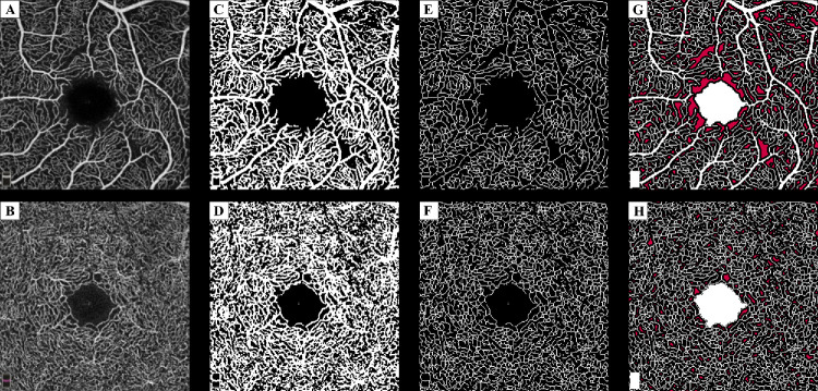
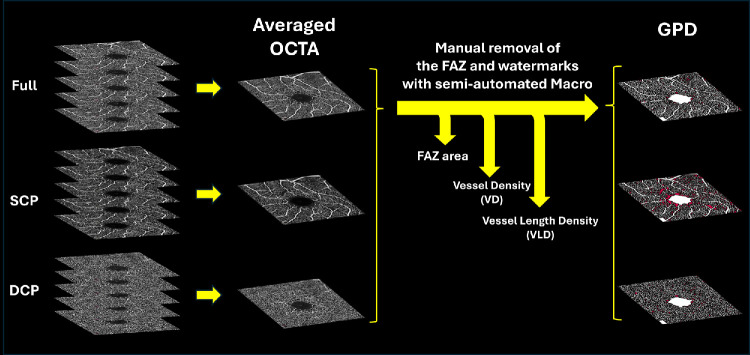
Methods: In this prospective, 1-year observational study, 208 patients with diabetes (320 eyes) underwent 3 × 3 mm macular OCTA at baseline, 6 months, and 12 months. Registered, averaged images yielded geometric perfusion deficits (GPDs), vessel density (VD), vessel length density (VLD) in the superficial and deep capillary plexuses (SCP and DCP), and foveal avascular zone area. Eyes were graded as non-referable or referable. Linear mixed models adjusted for age, sex, diabetes duration, hemoglobin A1c, and hypertension were conducted. Post hoc Dunnett's tests compared follow-ups with baseline within each severity group.
Results: A total of 709 eye visits were analyzed by OCTA. Across the cohort, referable DR, longer diabetes duration, and hypertension were independently associated with higher GPD values. In referable eyes, GPD-DCP increased at 6 months (P = 0.036) and 1 year (P = 0.016), whereas no other OCTA metric changed significantly at 6 months. In non-referable eyes, the only significant change was a decrease in VD-SCP at 1 year (P = 0.004).
Conclusions: Microvascular progression follows distinct, layer-specific patterns. In non-referable DR, capillary ischemia progresses more slowly and is more prominent in the SCP. In referable DR, GPD-DCP detected the earliest signs of microvascular progression and may serve as a promising biomarker, preceding vessel-based metrics. These findings suggest that the most informative OCTA metric should be tailored not only to the study question or timeline, but also to DR severity.
期刊介绍:
Investigative Ophthalmology & Visual Science (IOVS), published as ready online, is a peer-reviewed academic journal of the Association for Research in Vision and Ophthalmology (ARVO). IOVS features original research, mostly pertaining to clinical and laboratory ophthalmology and vision research in general.

 求助内容:
求助内容: 应助结果提醒方式:


