{"title":"一种新的小鼠颗粒状角膜营养不良II型模型揭示了自噬受损并概括了人类的发病机制。","authors":"Yanze Yu, Zhe Zhang, Zimeng Zhai, Bingqing Sun, Dongmei Yang, Zhanying Wang, Qinghong Lin, Xingtao Zhou, Jing Zhao","doi":"10.1167/iovs.66.12.7","DOIUrl":null,"url":null,"abstract":"<p><strong>Purpose: </strong>To develop and characterize a novel mouse model of granular corneal dystrophy type II (GCD2) using CRISPR/Cas9 technology and explore the underlying pathogenesis of transforming growth factor-beta-induced protein (TGFBIp) aggregation.</p><p><strong>Methods: </strong>CRISPR/Cas9 technology was employed to introduce the R124H mutation in the TGFBI gene of mice. Genomic sequencing and polymerase chain reaction confirmed the mutation. Phenotypic characteristics were evaluated through slit-lamp examination, optical coherence tomography, histological analysis, electron microscopy, and immunofluorescence, comparing wild-type (WT), heterozygous (HE), and homozygous (HO) mice. Transcriptome sequencing was conducted to identify the pathogenesis of GCD2. The findings were further validated through western blotting and transmission electron microscopy.</p><p><strong>Results: </strong>The R124H mutation in TGFBI was successfully introduced, with breadcrumb-like deposits observed in the corneas of mutant mice, with HO mice displaying more severe phenotypes than HE mice. TGFBIp levels were elevated in HE and HO mice (both P < 0.001). Histological and electron microscopy analyses revealed abnormal collagen arrangement and TGFBIp deposits in the corneal stroma of the HE and HO mice. Transcriptome analysis indicated that the TGFBI-R124H mutation was associated with impaired autophagy, endocytosis, and extracellular matrix signaling. Additional experiments confirmed autophagy-related markers LC3 and SQSTM1 were upregulated in the corneas of mutant mice, accompanied by increased autophagosome formation in corneal keratocytes, indicating impaired autophagy flux in HE and HO mice.</p><p><strong>Conclusions: </strong>We established a GCD2 mouse model caused by the R124H mutation using CRISPR/Cas9, providing a reliable platform for understanding pathogenesis for GCD2.</p>","PeriodicalId":14620,"journal":{"name":"Investigative ophthalmology & visual science","volume":"66 12","pages":"7"},"PeriodicalIF":4.7000,"publicationDate":"2025-09-02","publicationTypes":"Journal Article","fieldsOfStudy":null,"isOpenAccess":false,"openAccessPdf":"https://www.ncbi.nlm.nih.gov/pmc/articles/PMC12410266/pdf/","citationCount":"0","resultStr":"{\"title\":\"A Novel Mouse Model of Granular Corneal Dystrophy Type II Reveals Impaired Autophagy and Recapitulates Human Pathogenesis.\",\"authors\":\"Yanze Yu, Zhe Zhang, Zimeng Zhai, Bingqing Sun, Dongmei Yang, Zhanying Wang, Qinghong Lin, Xingtao Zhou, Jing Zhao\",\"doi\":\"10.1167/iovs.66.12.7\",\"DOIUrl\":null,\"url\":null,\"abstract\":\"<p><strong>Purpose: </strong>To develop and characterize a novel mouse model of granular corneal dystrophy type II (GCD2) using CRISPR/Cas9 technology and explore the underlying pathogenesis of transforming growth factor-beta-induced protein (TGFBIp) aggregation.</p><p><strong>Methods: </strong>CRISPR/Cas9 technology was employed to introduce the R124H mutation in the TGFBI gene of mice. Genomic sequencing and polymerase chain reaction confirmed the mutation. Phenotypic characteristics were evaluated through slit-lamp examination, optical coherence tomography, histological analysis, electron microscopy, and immunofluorescence, comparing wild-type (WT), heterozygous (HE), and homozygous (HO) mice. Transcriptome sequencing was conducted to identify the pathogenesis of GCD2. The findings were further validated through western blotting and transmission electron microscopy.</p><p><strong>Results: </strong>The R124H mutation in TGFBI was successfully introduced, with breadcrumb-like deposits observed in the corneas of mutant mice, with HO mice displaying more severe phenotypes than HE mice. TGFBIp levels were elevated in HE and HO mice (both P < 0.001). Histological and electron microscopy analyses revealed abnormal collagen arrangement and TGFBIp deposits in the corneal stroma of the HE and HO mice. Transcriptome analysis indicated that the TGFBI-R124H mutation was associated with impaired autophagy, endocytosis, and extracellular matrix signaling. Additional experiments confirmed autophagy-related markers LC3 and SQSTM1 were upregulated in the corneas of mutant mice, accompanied by increased autophagosome formation in corneal keratocytes, indicating impaired autophagy flux in HE and HO mice.</p><p><strong>Conclusions: </strong>We established a GCD2 mouse model caused by the R124H mutation using CRISPR/Cas9, providing a reliable platform for understanding pathogenesis for GCD2.</p>\",\"PeriodicalId\":14620,\"journal\":{\"name\":\"Investigative ophthalmology & visual science\",\"volume\":\"66 12\",\"pages\":\"7\"},\"PeriodicalIF\":4.7000,\"publicationDate\":\"2025-09-02\",\"publicationTypes\":\"Journal Article\",\"fieldsOfStudy\":null,\"isOpenAccess\":false,\"openAccessPdf\":\"https://www.ncbi.nlm.nih.gov/pmc/articles/PMC12410266/pdf/\",\"citationCount\":\"0\",\"resultStr\":null,\"platform\":\"Semanticscholar\",\"paperid\":null,\"PeriodicalName\":\"Investigative ophthalmology & visual science\",\"FirstCategoryId\":\"3\",\"ListUrlMain\":\"https://doi.org/10.1167/iovs.66.12.7\",\"RegionNum\":2,\"RegionCategory\":\"医学\",\"ArticlePicture\":[],\"TitleCN\":null,\"AbstractTextCN\":null,\"PMCID\":null,\"EPubDate\":\"\",\"PubModel\":\"\",\"JCR\":\"Q1\",\"JCRName\":\"OPHTHALMOLOGY\",\"Score\":null,\"Total\":0}","platform":"Semanticscholar","paperid":null,"PeriodicalName":"Investigative ophthalmology & visual science","FirstCategoryId":"3","ListUrlMain":"https://doi.org/10.1167/iovs.66.12.7","RegionNum":2,"RegionCategory":"医学","ArticlePicture":[],"TitleCN":null,"AbstractTextCN":null,"PMCID":null,"EPubDate":"","PubModel":"","JCR":"Q1","JCRName":"OPHTHALMOLOGY","Score":null,"Total":0}
A Novel Mouse Model of Granular Corneal Dystrophy Type II Reveals Impaired Autophagy and Recapitulates Human Pathogenesis.
Purpose: To develop and characterize a novel mouse model of granular corneal dystrophy type II (GCD2) using CRISPR/Cas9 technology and explore the underlying pathogenesis of transforming growth factor-beta-induced protein (TGFBIp) aggregation.
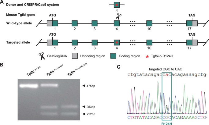
Methods: CRISPR/Cas9 technology was employed to introduce the R124H mutation in the TGFBI gene of mice. Genomic sequencing and polymerase chain reaction confirmed the mutation. Phenotypic characteristics were evaluated through slit-lamp examination, optical coherence tomography, histological analysis, electron microscopy, and immunofluorescence, comparing wild-type (WT), heterozygous (HE), and homozygous (HO) mice. Transcriptome sequencing was conducted to identify the pathogenesis of GCD2. The findings were further validated through western blotting and transmission electron microscopy.
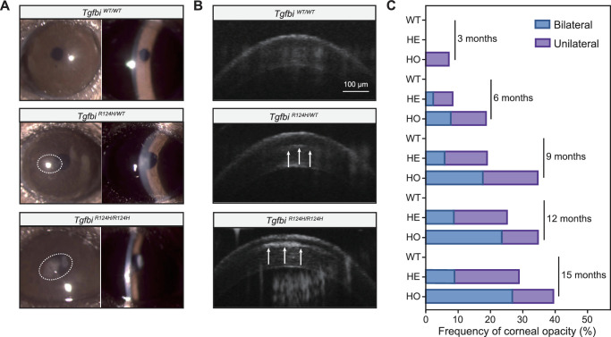
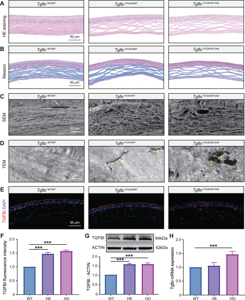
Results: The R124H mutation in TGFBI was successfully introduced, with breadcrumb-like deposits observed in the corneas of mutant mice, with HO mice displaying more severe phenotypes than HE mice. TGFBIp levels were elevated in HE and HO mice (both P < 0.001). Histological and electron microscopy analyses revealed abnormal collagen arrangement and TGFBIp deposits in the corneal stroma of the HE and HO mice. Transcriptome analysis indicated that the TGFBI-R124H mutation was associated with impaired autophagy, endocytosis, and extracellular matrix signaling. Additional experiments confirmed autophagy-related markers LC3 and SQSTM1 were upregulated in the corneas of mutant mice, accompanied by increased autophagosome formation in corneal keratocytes, indicating impaired autophagy flux in HE and HO mice.
Conclusions: We established a GCD2 mouse model caused by the R124H mutation using CRISPR/Cas9, providing a reliable platform for understanding pathogenesis for GCD2.
期刊介绍:
Investigative Ophthalmology & Visual Science (IOVS), published as ready online, is a peer-reviewed academic journal of the Association for Research in Vision and Ophthalmology (ARVO). IOVS features original research, mostly pertaining to clinical and laboratory ophthalmology and vision research in general.

 求助内容:
求助内容: 应助结果提醒方式:


