Liya Li, Ying Hu, Xiaojun Wang, Pei Sun, Tingwei Quan
{"title":"用于单神经元轴突重建的深度学习神经元成像数据集。","authors":"Liya Li, Ying Hu, Xiaojun Wang, Pei Sun, Tingwei Quan","doi":"10.3389/fninf.2025.1628030","DOIUrl":null,"url":null,"abstract":"<p><p>Neuron reconstruction is a critical step in quantifying neuronal structures from imaging data. Advances in molecular labeling techniques and optical imaging technologies have spurred extensive research into the patterns of long-range neuronal projections. However, mapping these projections incurs significant costs, as large-scale reconstruction of individual axonal arbors remains time-consuming. In this study, we present a dataset comprising axon imaging volumes along with corresponding annotations to facilitate the evaluation and development of axon reconstruction algorithms. This dataset, derived from 11 mouse brain samples imaged using fluorescence micro-optical sectioning tomography, contains carefully selected 852 volume images sized at 192 × 192 × 192 voxels. These images exhibit substantial variations in terms of axon density, image intensity, and signal-to-noise ratios, even within localized regions. Conventional methods often struggle when processing such complex data. To address these challenges, we propose a distance field-supervised segmentation network designed to enhance image signals effectively. Our results demonstrate significantly improved axon detection rates across both state-of-the-art and traditional methodologies. The released dataset and benchmark algorithm provide a data foundation for advancing novel axon reconstruction methods and are valuable for accelerating the reconstruction of long-range axonal projections.</p>","PeriodicalId":12462,"journal":{"name":"Frontiers in Neuroinformatics","volume":"19 ","pages":"1628030"},"PeriodicalIF":2.5000,"publicationDate":"2025-08-05","publicationTypes":"Journal Article","fieldsOfStudy":null,"isOpenAccess":false,"openAccessPdf":"https://www.ncbi.nlm.nih.gov/pmc/articles/PMC12361129/pdf/","citationCount":"0","resultStr":"{\"title\":\"A neuronal imaging dataset for deep learning in the reconstruction of single-neuron axons.\",\"authors\":\"Liya Li, Ying Hu, Xiaojun Wang, Pei Sun, Tingwei Quan\",\"doi\":\"10.3389/fninf.2025.1628030\",\"DOIUrl\":null,\"url\":null,\"abstract\":\"<p><p>Neuron reconstruction is a critical step in quantifying neuronal structures from imaging data. Advances in molecular labeling techniques and optical imaging technologies have spurred extensive research into the patterns of long-range neuronal projections. However, mapping these projections incurs significant costs, as large-scale reconstruction of individual axonal arbors remains time-consuming. In this study, we present a dataset comprising axon imaging volumes along with corresponding annotations to facilitate the evaluation and development of axon reconstruction algorithms. This dataset, derived from 11 mouse brain samples imaged using fluorescence micro-optical sectioning tomography, contains carefully selected 852 volume images sized at 192 × 192 × 192 voxels. These images exhibit substantial variations in terms of axon density, image intensity, and signal-to-noise ratios, even within localized regions. Conventional methods often struggle when processing such complex data. To address these challenges, we propose a distance field-supervised segmentation network designed to enhance image signals effectively. Our results demonstrate significantly improved axon detection rates across both state-of-the-art and traditional methodologies. The released dataset and benchmark algorithm provide a data foundation for advancing novel axon reconstruction methods and are valuable for accelerating the reconstruction of long-range axonal projections.</p>\",\"PeriodicalId\":12462,\"journal\":{\"name\":\"Frontiers in Neuroinformatics\",\"volume\":\"19 \",\"pages\":\"1628030\"},\"PeriodicalIF\":2.5000,\"publicationDate\":\"2025-08-05\",\"publicationTypes\":\"Journal Article\",\"fieldsOfStudy\":null,\"isOpenAccess\":false,\"openAccessPdf\":\"https://www.ncbi.nlm.nih.gov/pmc/articles/PMC12361129/pdf/\",\"citationCount\":\"0\",\"resultStr\":null,\"platform\":\"Semanticscholar\",\"paperid\":null,\"PeriodicalName\":\"Frontiers in Neuroinformatics\",\"FirstCategoryId\":\"3\",\"ListUrlMain\":\"https://doi.org/10.3389/fninf.2025.1628030\",\"RegionNum\":4,\"RegionCategory\":\"医学\",\"ArticlePicture\":[],\"TitleCN\":null,\"AbstractTextCN\":null,\"PMCID\":null,\"EPubDate\":\"2025/1/1 0:00:00\",\"PubModel\":\"eCollection\",\"JCR\":\"Q2\",\"JCRName\":\"MATHEMATICAL & COMPUTATIONAL BIOLOGY\",\"Score\":null,\"Total\":0}","platform":"Semanticscholar","paperid":null,"PeriodicalName":"Frontiers in Neuroinformatics","FirstCategoryId":"3","ListUrlMain":"https://doi.org/10.3389/fninf.2025.1628030","RegionNum":4,"RegionCategory":"医学","ArticlePicture":[],"TitleCN":null,"AbstractTextCN":null,"PMCID":null,"EPubDate":"2025/1/1 0:00:00","PubModel":"eCollection","JCR":"Q2","JCRName":"MATHEMATICAL & COMPUTATIONAL BIOLOGY","Score":null,"Total":0}
A neuronal imaging dataset for deep learning in the reconstruction of single-neuron axons.
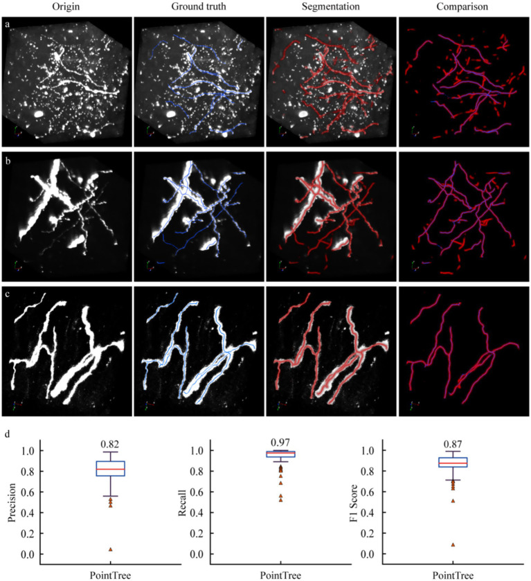
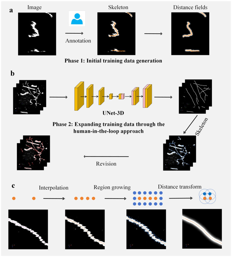
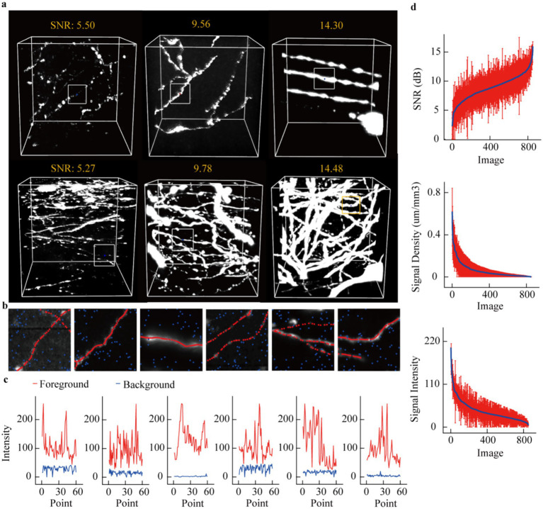
Neuron reconstruction is a critical step in quantifying neuronal structures from imaging data. Advances in molecular labeling techniques and optical imaging technologies have spurred extensive research into the patterns of long-range neuronal projections. However, mapping these projections incurs significant costs, as large-scale reconstruction of individual axonal arbors remains time-consuming. In this study, we present a dataset comprising axon imaging volumes along with corresponding annotations to facilitate the evaluation and development of axon reconstruction algorithms. This dataset, derived from 11 mouse brain samples imaged using fluorescence micro-optical sectioning tomography, contains carefully selected 852 volume images sized at 192 × 192 × 192 voxels. These images exhibit substantial variations in terms of axon density, image intensity, and signal-to-noise ratios, even within localized regions. Conventional methods often struggle when processing such complex data. To address these challenges, we propose a distance field-supervised segmentation network designed to enhance image signals effectively. Our results demonstrate significantly improved axon detection rates across both state-of-the-art and traditional methodologies. The released dataset and benchmark algorithm provide a data foundation for advancing novel axon reconstruction methods and are valuable for accelerating the reconstruction of long-range axonal projections.
期刊介绍:
Frontiers in Neuroinformatics publishes rigorously peer-reviewed research on the development and implementation of numerical/computational models and analytical tools used to share, integrate and analyze experimental data and advance theories of the nervous system functions. Specialty Chief Editors Jan G. Bjaalie at the University of Oslo and Sean L. Hill at the École Polytechnique Fédérale de Lausanne are supported by an outstanding Editorial Board of international experts. This multidisciplinary open-access journal is at the forefront of disseminating and communicating scientific knowledge and impactful discoveries to researchers, academics and the public worldwide.
Neuroscience is being propelled into the information age as the volume of information explodes, demanding organization and synthesis. Novel synthesis approaches are opening up a new dimension for the exploration of the components of brain elements and systems and the vast number of variables that underlie their functions. Neural data is highly heterogeneous with complex inter-relations across multiple levels, driving the need for innovative organizing and synthesizing approaches from genes to cognition, and covering a range of species and disease states.
Frontiers in Neuroinformatics therefore welcomes submissions on existing neuroscience databases, development of data and knowledge bases for all levels of neuroscience, applications and technologies that can facilitate data sharing (interoperability, formats, terminologies, and ontologies), and novel tools for data acquisition, analyses, visualization, and dissemination of nervous system data. Our journal welcomes submissions on new tools (software and hardware) that support brain modeling, and the merging of neuroscience databases with brain models used for simulation and visualization.
 求助内容:
求助内容: 应助结果提醒方式:


