{"title":"青光眼患者黄斑色素光学体积与视功能的关系。","authors":"Norikazu Matsumura, Ryo Asaoka, Yuri Fujino, Akira Obana","doi":"10.1167/iovs.66.11.31","DOIUrl":null,"url":null,"abstract":"<p><strong>Purpose: </strong>This study aimed to investigate the relationships among visual field (VF) sensitivity, retinal structure, and macular pigment in glaucoma.</p><p><strong>Methods: </strong>A total of 218 eyes from 121 patients diagnosed with primary open-angle glaucoma were included. VF sensitivity was assessed using the 10-2 test with the Humphrey Field Analyzer. Macular pigment optical volume (MPOV) was measured using the two-wavelength fundus autofluorescence method with SPECTRALIS optical coherence tomography (OCT). Additionally, ganglion cell complex (GCC) and nerve fiber layer (NFL) thickness were evaluated using OCT. The relationships among VF sensitivity, retinal structure, and MPOV were evaluated by matching the measurement areas of each test as closely as possible.</p><p><strong>Results: </strong>The mean age of participants was 71.6 ± 10.8 years (49 males). MPOV showed no significant association with VF sensitivity in either the superior or inferior hemiretina (P = 0.75 and P = 0.42, respectively). MPOV was not significantly associated with GCC or NFL thickness in both the superior and inferior hemiretina (P > 0.05).</p><p><strong>Conclusions: </strong>In glaucoma patients, MPOV was not associated with VF sensitivity, GCC, or NFL. These findings suggest no structural or functional association between MPOV and glaucomatous damage.</p>","PeriodicalId":14620,"journal":{"name":"Investigative ophthalmology & visual science","volume":"66 11","pages":"31"},"PeriodicalIF":4.7000,"publicationDate":"2025-08-01","publicationTypes":"Journal Article","fieldsOfStudy":null,"isOpenAccess":false,"openAccessPdf":"https://www.ncbi.nlm.nih.gov/pmc/articles/PMC12369915/pdf/","citationCount":"0","resultStr":"{\"title\":\"The Relationship Between Macular Pigment Optical Volume and Visual Function in Glaucoma Patients.\",\"authors\":\"Norikazu Matsumura, Ryo Asaoka, Yuri Fujino, Akira Obana\",\"doi\":\"10.1167/iovs.66.11.31\",\"DOIUrl\":null,\"url\":null,\"abstract\":\"<p><strong>Purpose: </strong>This study aimed to investigate the relationships among visual field (VF) sensitivity, retinal structure, and macular pigment in glaucoma.</p><p><strong>Methods: </strong>A total of 218 eyes from 121 patients diagnosed with primary open-angle glaucoma were included. VF sensitivity was assessed using the 10-2 test with the Humphrey Field Analyzer. Macular pigment optical volume (MPOV) was measured using the two-wavelength fundus autofluorescence method with SPECTRALIS optical coherence tomography (OCT). Additionally, ganglion cell complex (GCC) and nerve fiber layer (NFL) thickness were evaluated using OCT. The relationships among VF sensitivity, retinal structure, and MPOV were evaluated by matching the measurement areas of each test as closely as possible.</p><p><strong>Results: </strong>The mean age of participants was 71.6 ± 10.8 years (49 males). MPOV showed no significant association with VF sensitivity in either the superior or inferior hemiretina (P = 0.75 and P = 0.42, respectively). MPOV was not significantly associated with GCC or NFL thickness in both the superior and inferior hemiretina (P > 0.05).</p><p><strong>Conclusions: </strong>In glaucoma patients, MPOV was not associated with VF sensitivity, GCC, or NFL. These findings suggest no structural or functional association between MPOV and glaucomatous damage.</p>\",\"PeriodicalId\":14620,\"journal\":{\"name\":\"Investigative ophthalmology & visual science\",\"volume\":\"66 11\",\"pages\":\"31\"},\"PeriodicalIF\":4.7000,\"publicationDate\":\"2025-08-01\",\"publicationTypes\":\"Journal Article\",\"fieldsOfStudy\":null,\"isOpenAccess\":false,\"openAccessPdf\":\"https://www.ncbi.nlm.nih.gov/pmc/articles/PMC12369915/pdf/\",\"citationCount\":\"0\",\"resultStr\":null,\"platform\":\"Semanticscholar\",\"paperid\":null,\"PeriodicalName\":\"Investigative ophthalmology & visual science\",\"FirstCategoryId\":\"3\",\"ListUrlMain\":\"https://doi.org/10.1167/iovs.66.11.31\",\"RegionNum\":2,\"RegionCategory\":\"医学\",\"ArticlePicture\":[],\"TitleCN\":null,\"AbstractTextCN\":null,\"PMCID\":null,\"EPubDate\":\"\",\"PubModel\":\"\",\"JCR\":\"Q1\",\"JCRName\":\"OPHTHALMOLOGY\",\"Score\":null,\"Total\":0}","platform":"Semanticscholar","paperid":null,"PeriodicalName":"Investigative ophthalmology & visual science","FirstCategoryId":"3","ListUrlMain":"https://doi.org/10.1167/iovs.66.11.31","RegionNum":2,"RegionCategory":"医学","ArticlePicture":[],"TitleCN":null,"AbstractTextCN":null,"PMCID":null,"EPubDate":"","PubModel":"","JCR":"Q1","JCRName":"OPHTHALMOLOGY","Score":null,"Total":0}
The Relationship Between Macular Pigment Optical Volume and Visual Function in Glaucoma Patients.
Purpose: This study aimed to investigate the relationships among visual field (VF) sensitivity, retinal structure, and macular pigment in glaucoma.
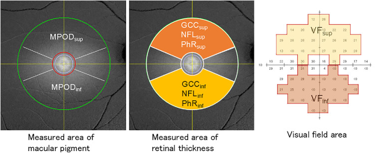
Methods: A total of 218 eyes from 121 patients diagnosed with primary open-angle glaucoma were included. VF sensitivity was assessed using the 10-2 test with the Humphrey Field Analyzer. Macular pigment optical volume (MPOV) was measured using the two-wavelength fundus autofluorescence method with SPECTRALIS optical coherence tomography (OCT). Additionally, ganglion cell complex (GCC) and nerve fiber layer (NFL) thickness were evaluated using OCT. The relationships among VF sensitivity, retinal structure, and MPOV were evaluated by matching the measurement areas of each test as closely as possible.
Results: The mean age of participants was 71.6 ± 10.8 years (49 males). MPOV showed no significant association with VF sensitivity in either the superior or inferior hemiretina (P = 0.75 and P = 0.42, respectively). MPOV was not significantly associated with GCC or NFL thickness in both the superior and inferior hemiretina (P > 0.05).
Conclusions: In glaucoma patients, MPOV was not associated with VF sensitivity, GCC, or NFL. These findings suggest no structural or functional association between MPOV and glaucomatous damage.
期刊介绍:
Investigative Ophthalmology & Visual Science (IOVS), published as ready online, is a peer-reviewed academic journal of the Association for Research in Vision and Ophthalmology (ARVO). IOVS features original research, mostly pertaining to clinical and laboratory ophthalmology and vision research in general.

 求助内容:
求助内容: 应助结果提醒方式:


