Panagiotis N Toumasis, Ashwin Mallipatna, Timothy W Corson, Helen Dimaras
{"title":"视网膜瘤:综述。","authors":"Panagiotis N Toumasis, Ashwin Mallipatna, Timothy W Corson, Helen Dimaras","doi":"10.1002/ped4.12472","DOIUrl":null,"url":null,"abstract":"<p><p>Retinoma, also referred to as retinocytoma, is a benign manifestation of biallelic retinoblastoma gene (<i>RB1</i>) inactivation. Genetic or epigenetic loss of retinoblastoma protein in maturing cone precursors induces genomic instability which leads to upregulation of senescence-associated p16<sup>INK4a</sup> and p130, resulting in non-proliferative retinoma. When senescence pathways fail and genetic instability accumulates to a critical level through altered gene copies of oncogenes and tumor suppression genes, transformation into <i>RB1</i> <sup>-/-</sup> retinoblastoma occurs. Thus, the management of retinoma involves frequent ophthalmic examination and imaging to monitor the size and characteristics of the tumor, ensure stability, and rule out malignant transformation. Key ophthalmoscopic features of retinoma often include a translucent whitish-gray retinal mass, calcification, retinal pigment epithelial alterations with well-defined margins, located typically around the lesion, as well as a zone of chorioretinal atrophy. This review aims to provide a comprehensive overview of this non-malignant tumor drawing from current understanding of its molecular genetics, clinical characteristics, diagnostic modalities, differential diagnosis, management, and prognosis. A deeper understanding of retinoma could offer valuable insights into how retinoblastoma develops and oncogenesis more broadly, paving the way for improved strategies to prevent and treat this malignant tumor.</p>","PeriodicalId":19992,"journal":{"name":"Pediatric Investigation","volume":"9 2","pages":"139-149"},"PeriodicalIF":2.0000,"publicationDate":"2025-03-10","publicationTypes":"Journal Article","fieldsOfStudy":null,"isOpenAccess":false,"openAccessPdf":"https://www.ncbi.nlm.nih.gov/pmc/articles/PMC12175641/pdf/","citationCount":"0","resultStr":"{\"title\":\"Retinoma: An overview.\",\"authors\":\"Panagiotis N Toumasis, Ashwin Mallipatna, Timothy W Corson, Helen Dimaras\",\"doi\":\"10.1002/ped4.12472\",\"DOIUrl\":null,\"url\":null,\"abstract\":\"<p><p>Retinoma, also referred to as retinocytoma, is a benign manifestation of biallelic retinoblastoma gene (<i>RB1</i>) inactivation. Genetic or epigenetic loss of retinoblastoma protein in maturing cone precursors induces genomic instability which leads to upregulation of senescence-associated p16<sup>INK4a</sup> and p130, resulting in non-proliferative retinoma. When senescence pathways fail and genetic instability accumulates to a critical level through altered gene copies of oncogenes and tumor suppression genes, transformation into <i>RB1</i> <sup>-/-</sup> retinoblastoma occurs. Thus, the management of retinoma involves frequent ophthalmic examination and imaging to monitor the size and characteristics of the tumor, ensure stability, and rule out malignant transformation. Key ophthalmoscopic features of retinoma often include a translucent whitish-gray retinal mass, calcification, retinal pigment epithelial alterations with well-defined margins, located typically around the lesion, as well as a zone of chorioretinal atrophy. This review aims to provide a comprehensive overview of this non-malignant tumor drawing from current understanding of its molecular genetics, clinical characteristics, diagnostic modalities, differential diagnosis, management, and prognosis. A deeper understanding of retinoma could offer valuable insights into how retinoblastoma develops and oncogenesis more broadly, paving the way for improved strategies to prevent and treat this malignant tumor.</p>\",\"PeriodicalId\":19992,\"journal\":{\"name\":\"Pediatric Investigation\",\"volume\":\"9 2\",\"pages\":\"139-149\"},\"PeriodicalIF\":2.0000,\"publicationDate\":\"2025-03-10\",\"publicationTypes\":\"Journal Article\",\"fieldsOfStudy\":null,\"isOpenAccess\":false,\"openAccessPdf\":\"https://www.ncbi.nlm.nih.gov/pmc/articles/PMC12175641/pdf/\",\"citationCount\":\"0\",\"resultStr\":null,\"platform\":\"Semanticscholar\",\"paperid\":null,\"PeriodicalName\":\"Pediatric Investigation\",\"FirstCategoryId\":\"3\",\"ListUrlMain\":\"https://doi.org/10.1002/ped4.12472\",\"RegionNum\":4,\"RegionCategory\":\"医学\",\"ArticlePicture\":[],\"TitleCN\":null,\"AbstractTextCN\":null,\"PMCID\":null,\"EPubDate\":\"2025/6/1 0:00:00\",\"PubModel\":\"eCollection\",\"JCR\":\"Q2\",\"JCRName\":\"PEDIATRICS\",\"Score\":null,\"Total\":0}","platform":"Semanticscholar","paperid":null,"PeriodicalName":"Pediatric Investigation","FirstCategoryId":"3","ListUrlMain":"https://doi.org/10.1002/ped4.12472","RegionNum":4,"RegionCategory":"医学","ArticlePicture":[],"TitleCN":null,"AbstractTextCN":null,"PMCID":null,"EPubDate":"2025/6/1 0:00:00","PubModel":"eCollection","JCR":"Q2","JCRName":"PEDIATRICS","Score":null,"Total":0}
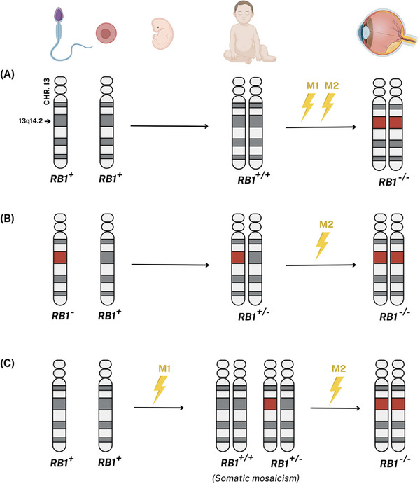
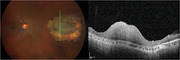
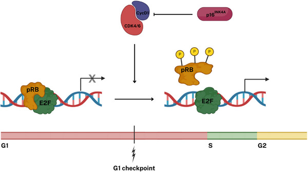
Retinoma, also referred to as retinocytoma, is a benign manifestation of biallelic retinoblastoma gene (RB1) inactivation. Genetic or epigenetic loss of retinoblastoma protein in maturing cone precursors induces genomic instability which leads to upregulation of senescence-associated p16INK4a and p130, resulting in non-proliferative retinoma. When senescence pathways fail and genetic instability accumulates to a critical level through altered gene copies of oncogenes and tumor suppression genes, transformation into RB1-/- retinoblastoma occurs. Thus, the management of retinoma involves frequent ophthalmic examination and imaging to monitor the size and characteristics of the tumor, ensure stability, and rule out malignant transformation. Key ophthalmoscopic features of retinoma often include a translucent whitish-gray retinal mass, calcification, retinal pigment epithelial alterations with well-defined margins, located typically around the lesion, as well as a zone of chorioretinal atrophy. This review aims to provide a comprehensive overview of this non-malignant tumor drawing from current understanding of its molecular genetics, clinical characteristics, diagnostic modalities, differential diagnosis, management, and prognosis. A deeper understanding of retinoma could offer valuable insights into how retinoblastoma develops and oncogenesis more broadly, paving the way for improved strategies to prevent and treat this malignant tumor.
 求助内容:
求助内容: 应助结果提醒方式:


