基于可编程高光谱成像的无标签眼底血管造影术

IF 4.6 2区 物理与天体物理 Q1 OPTICS
Yuetian Ren , Yuning Song , Jinchi Yao , Hongguo Wei , Yunxin Wang , Shuaihang Luo , Wenbin Xu , James Joseph , Keren Chen , Shuo Chen
{"title":"基于可编程高光谱成像的无标签眼底血管造影术","authors":"Yuetian Ren ,&nbsp;Yuning Song ,&nbsp;Jinchi Yao ,&nbsp;Hongguo Wei ,&nbsp;Yunxin Wang ,&nbsp;Shuaihang Luo ,&nbsp;Wenbin Xu ,&nbsp;James Joseph ,&nbsp;Keren Chen ,&nbsp;Shuo Chen","doi":"10.1016/j.optlastec.2025.113389","DOIUrl":null,"url":null,"abstract":"<div><div>Retinal vasculature, as the sole deep vascular network within the human body that can be non-invasively observed, is closely associated with various retinal and systemic disorders. However, conventional fundus imaging techniques often suffer from inadequate contrast or rely on invasive contrast agents to visualize fine retinal vessels, hemorrhages, and exudates, thereby posing great challenges in screening or diagnosing fundus diseases at their early stages. In this study, we report a label-free optical fundus angiography method based on programmable hyperspectral imaging, in which fundus images with high-contrast retinal blood vessels can be captured in a snapshot by exposing fundus tissues under specific spectrally coded illumination. Using a self-developed programmable light source, such specific spectrally coded illumination is multiplexed with specific intensities at different wavelengths under the guidance of spectral post-processing algorithms. This enables the preservation of key spectral bands that can maximize the differences between the retinal blood vessels and other fundus tissues. In such a manner, the proposed method can select the wavelengths of interest during fundus imaging in a snapshot, in contrast to numerical wavelength selection after acquiring the full wavelengths of conventional spectroscopic fundus imaging techniques; thus, the time-consuming acquisition of a three-dimensional spectral dataset and spectral post-processing can be avoided. Through evaluations on a model eye and ex vivo porcine eyes, the proposed label-free optical fundus angiography method shows significant enhancement in the image contrast of retinal blood vessels compared to conventional fundus imaging, thus is expected to benefit early and more precise diagnosis of fundus diseases.</div></div>","PeriodicalId":19511,"journal":{"name":"Optics and Laser Technology","volume":"191 ","pages":"Article 113389"},"PeriodicalIF":4.6000,"publicationDate":"2025-06-16","publicationTypes":"Journal Article","fieldsOfStudy":null,"isOpenAccess":false,"openAccessPdf":"","citationCount":"0","resultStr":"{\"title\":\"Label-free optical fundus angiography based on programmable hyperspectral imaging\",\"authors\":\"Yuetian Ren ,&nbsp;Yuning Song ,&nbsp;Jinchi Yao ,&nbsp;Hongguo Wei ,&nbsp;Yunxin Wang ,&nbsp;Shuaihang Luo ,&nbsp;Wenbin Xu ,&nbsp;James Joseph ,&nbsp;Keren Chen ,&nbsp;Shuo Chen\",\"doi\":\"10.1016/j.optlastec.2025.113389\",\"DOIUrl\":null,\"url\":null,\"abstract\":\"<div><div>Retinal vasculature, as the sole deep vascular network within the human body that can be non-invasively observed, is closely associated with various retinal and systemic disorders. However, conventional fundus imaging techniques often suffer from inadequate contrast or rely on invasive contrast agents to visualize fine retinal vessels, hemorrhages, and exudates, thereby posing great challenges in screening or diagnosing fundus diseases at their early stages. In this study, we report a label-free optical fundus angiography method based on programmable hyperspectral imaging, in which fundus images with high-contrast retinal blood vessels can be captured in a snapshot by exposing fundus tissues under specific spectrally coded illumination. Using a self-developed programmable light source, such specific spectrally coded illumination is multiplexed with specific intensities at different wavelengths under the guidance of spectral post-processing algorithms. This enables the preservation of key spectral bands that can maximize the differences between the retinal blood vessels and other fundus tissues. In such a manner, the proposed method can select the wavelengths of interest during fundus imaging in a snapshot, in contrast to numerical wavelength selection after acquiring the full wavelengths of conventional spectroscopic fundus imaging techniques; thus, the time-consuming acquisition of a three-dimensional spectral dataset and spectral post-processing can be avoided. Through evaluations on a model eye and ex vivo porcine eyes, the proposed label-free optical fundus angiography method shows significant enhancement in the image contrast of retinal blood vessels compared to conventional fundus imaging, thus is expected to benefit early and more precise diagnosis of fundus diseases.</div></div>\",\"PeriodicalId\":19511,\"journal\":{\"name\":\"Optics and Laser Technology\",\"volume\":\"191 \",\"pages\":\"Article 113389\"},\"PeriodicalIF\":4.6000,\"publicationDate\":\"2025-06-16\",\"publicationTypes\":\"Journal Article\",\"fieldsOfStudy\":null,\"isOpenAccess\":false,\"openAccessPdf\":\"\",\"citationCount\":\"0\",\"resultStr\":null,\"platform\":\"Semanticscholar\",\"paperid\":null,\"PeriodicalName\":\"Optics and Laser Technology\",\"FirstCategoryId\":\"101\",\"ListUrlMain\":\"https://www.sciencedirect.com/science/article/pii/S0030399225009806\",\"RegionNum\":2,\"RegionCategory\":\"物理与天体物理\",\"ArticlePicture\":[],\"TitleCN\":null,\"AbstractTextCN\":null,\"PMCID\":null,\"EPubDate\":\"\",\"PubModel\":\"\",\"JCR\":\"Q1\",\"JCRName\":\"OPTICS\",\"Score\":null,\"Total\":0}","platform":"Semanticscholar","paperid":null,"PeriodicalName":"Optics and Laser Technology","FirstCategoryId":"101","ListUrlMain":"https://www.sciencedirect.com/science/article/pii/S0030399225009806","RegionNum":2,"RegionCategory":"物理与天体物理","ArticlePicture":[],"TitleCN":null,"AbstractTextCN":null,"PMCID":null,"EPubDate":"","PubModel":"","JCR":"Q1","JCRName":"OPTICS","Score":null,"Total":0}
引用次数: 0

摘要

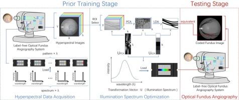
视网膜脉管系统作为人体内唯一可以无创观察到的深层血管网络,与各种视网膜及全身疾病密切相关。然而,传统的眼底成像技术往往存在对比度不足或依赖侵入性造影剂来显示精细视网膜血管、出血和渗出物的问题,因此在早期筛查或诊断眼底疾病时面临很大挑战。在这项研究中,我们报告了一种基于可编程高光谱成像的无标签光学眼底血管成像方法,该方法通过在特定的光谱编码照明下暴露眼底组织,可以在快照中捕获具有高对比度视网膜血管的眼底图像。利用自主开发的可编程光源,在光谱后处理算法的指导下,将特定的光谱编码照明与不同波长的特定强度进行复用。这使得保存关键的光谱带,可以最大限度地区分视网膜血管和其他眼底组织。通过这种方式,与传统光谱眼底成像技术在获得全波长后进行数值波长选择相比,该方法可以在快照中选择眼底成像过程中感兴趣的波长;因此,可以避免耗时的三维光谱数据采集和光谱后处理。通过对模型眼和离体猪眼的评估,所提出的无标记光学眼底血管造影方法与常规眼底成像相比,视网膜血管的图像对比度明显增强,有望更早、更准确地诊断眼底疾病。
本文章由计算机程序翻译,如有差异,请以英文原文为准。
Label-free optical fundus angiography based on programmable hyperspectral imaging
Retinal vasculature, as the sole deep vascular network within the human body that can be non-invasively observed, is closely associated with various retinal and systemic disorders. However, conventional fundus imaging techniques often suffer from inadequate contrast or rely on invasive contrast agents to visualize fine retinal vessels, hemorrhages, and exudates, thereby posing great challenges in screening or diagnosing fundus diseases at their early stages. In this study, we report a label-free optical fundus angiography method based on programmable hyperspectral imaging, in which fundus images with high-contrast retinal blood vessels can be captured in a snapshot by exposing fundus tissues under specific spectrally coded illumination. Using a self-developed programmable light source, such specific spectrally coded illumination is multiplexed with specific intensities at different wavelengths under the guidance of spectral post-processing algorithms. This enables the preservation of key spectral bands that can maximize the differences between the retinal blood vessels and other fundus tissues. In such a manner, the proposed method can select the wavelengths of interest during fundus imaging in a snapshot, in contrast to numerical wavelength selection after acquiring the full wavelengths of conventional spectroscopic fundus imaging techniques; thus, the time-consuming acquisition of a three-dimensional spectral dataset and spectral post-processing can be avoided. Through evaluations on a model eye and ex vivo porcine eyes, the proposed label-free optical fundus angiography method shows significant enhancement in the image contrast of retinal blood vessels compared to conventional fundus imaging, thus is expected to benefit early and more precise diagnosis of fundus diseases.
求助全文
通过发布文献求助,成功后即可免费获取论文全文。 去求助
来源期刊
CiteScore
8.50
自引率
10.00%
发文量
1060
审稿时长
3.4 months
期刊介绍: Optics & Laser Technology aims to provide a vehicle for the publication of a broad range of high quality research and review papers in those fields of scientific and engineering research appertaining to the development and application of the technology of optics and lasers. Papers describing original work in these areas are submitted to rigorous refereeing prior to acceptance for publication. The scope of Optics & Laser Technology encompasses, but is not restricted to, the following areas: •development in all types of lasers •developments in optoelectronic devices and photonics •developments in new photonics and optical concepts •developments in conventional optics, optical instruments and components •techniques of optical metrology, including interferometry and optical fibre sensors •LIDAR and other non-contact optical measurement techniques, including optical methods in heat and fluid flow •applications of lasers to materials processing, optical NDT display (including holography) and optical communication •research and development in the field of laser safety including studies of hazards resulting from the applications of lasers (laser safety, hazards of laser fume) •developments in optical computing and optical information processing •developments in new optical materials •developments in new optical characterization methods and techniques •developments in quantum optics •developments in light assisted micro and nanofabrication methods and techniques •developments in nanophotonics and biophotonics •developments in imaging processing and systems
×
引用
GB/T 7714-2015
复制
MLA
复制
APA
复制
导出至
BibTeX EndNote RefMan NoteFirst NoteExpress
×
提示
您的信息不完整,为了账户安全,请先补充。
现在去补充
×
提示
您因"违规操作"
具体请查看互助需知
我知道了
×
提示
确定
请完成安全验证×
copy
已复制链接
快去分享给好友吧!
我知道了
右上角分享
点击右上角分享
0
联系我们:info@booksci.cn Book学术提供免费学术资源搜索服务,方便国内外学者检索中英文文献。致力于提供最便捷和优质的服务体验。 Copyright © 2023 布克学术 All rights reserved.
京ICP备2023020795号-1
ghs 京公网安备 11010802042870号
Book学术文献互助
Book学术文献互助群
群 号:604180095
Book学术官方微信