甲下表皮样包涵体——附8例报告及文献复习。
IF 1.1
4区 医学
Q3 DERMATOLOGY
引用次数: 0
摘要
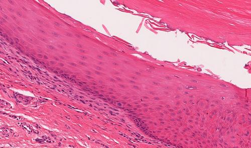
甲下表皮样包涵体(SEI)是甲床的良性囊性病变。据我们所知,只有一个案例系列描述了SEI。我们报告8例SEI。患者中位年龄为72岁(范围3-84岁),男女比例为1.6。5例发生在脚趾甲,3例发生在手指甲。组织学上,SEI的特征是网状嵴的球根状增生和单眼囊肿,内衬薄鳞状上皮,颗粒减少,充满正角蛋白。与甲床上皮的连接可能被破坏和钙化。趾下甲床病变的鉴别诊断应保留SEI。本文章由计算机程序翻译,如有差异,请以英文原文为准。
Subungual Epidermoid Inclusions–A Series of 8 Cases and a Review of Literature
Subungual epidermoid inclusions (SEI) are benign cystic lesions of the nail bed. To our knowledge, there has been only one case series describing SEI. We report eight cases of SEI. The patients had a median age of 72 years (range 3–84) with a female: male ratio of 1.6. Five occurred in toenails and three in fingernails. Histologically, SEI is characterized by bulbous proliferation of rete ridges and unilocular cysts lined by thin squamous epithelium with hypogranulosis, filled with orthokeratin. The connection to the nail bed epithelium may be disrupted and calcified. SEI are tumors that should be kept in the differential diagnosis of the subungual nail bed lesions.
求助全文
通过发布文献求助,成功后即可免费获取论文全文。
去求助
来源期刊

CiteScore
3.20
自引率
5.90%
发文量
174
审稿时长
3-8 weeks
期刊介绍:
Journal of Cutaneous Pathology publishes manuscripts broadly relevant to diseases of the skin and mucosae, with the aims of advancing scientific knowledge regarding dermatopathology and enhancing the communication between clinical practitioners and research scientists. Original scientific manuscripts on diagnostic and experimental cutaneous pathology are especially desirable. Timely, pertinent review articles also will be given high priority. Manuscripts based on light, fluorescence, and electron microscopy, histochemistry, immunology, molecular biology, and genetics, as well as allied sciences, are all welcome, provided their principal focus is on cutaneous pathology. Publication time will be kept as short as possible, ensuring that articles will be quickly available to all interested in this speciality.
 求助内容:
求助内容: 应助结果提醒方式:


