{"title":"磁共振波谱高琥珀酸峰:琥珀酸脱氢酶缺乏所致脑白质病的诊断线索。","authors":"Habibe Koc Ucar, Leman Tekin Orgun, Ebru Arhan, Ayse Serdaroglu, Kursad Aydin","doi":"10.22037/ijcn.v19i1.35156","DOIUrl":null,"url":null,"abstract":"<p><p>The Succinate Dehydrogenase (SDH) enzyme is known as Complex-II in the electron transport chain. This study reports the clinical and molecular investigations of three pediatric patients (two of whom are siblings), with histochemical and biochemical evidence of a severe, isolated complex II deficiency due to SDH gene mutations. The patients presented with severe hypotonia, developmental delay, spasticity, macrocephaly, and megalencephaly. Magnetic Resonance Imaging (MRI) revealed signal changes in the frontal, temporal, parietal, occipital cerebral, and cerebellar white matter, corpus striatum, thalamus, substantia nigra, inferior olivary nucleus, pyramidal tracts at the level of the pons and posterior limb of the internal capsule. Other typical findings involved a high succinate peak at 2.42 ppm and lactate peak at 1.3 ppm in Magnetic Resonance Spectroscopy (MRS). The siblings presented due to compound heterozygous c.143A>T (p. Asp48Val) and c.308T>C (p. Met103Thr) SDHB mutations, while the other patient presented due to compound heterozygous c.1754G>A (p. Arg585Gln) and c.1786G>C (p. Asp596His) SDHA mutation. The demonstration of succinate peak, particularly MRS, is highly diagnostic regarding SDH deficiency. MRS should be a standard part of routine radiological exams when there is a suspicion of a neurometabolic disease, especially mitochondrial disorders. Additionally, employing Next-Generation Sequencing (NGS) is advisable for patients as it allows for accurate diagnosis without requiring invasive procedures like muscle biopsies.</p>","PeriodicalId":14537,"journal":{"name":"Iranian Journal of Child Neurology","volume":"19 1","pages":"97-105"},"PeriodicalIF":0.9000,"publicationDate":"2025-01-01","publicationTypes":"Journal Article","fieldsOfStudy":null,"isOpenAccess":false,"openAccessPdf":"https://www.ncbi.nlm.nih.gov/pmc/articles/PMC11781339/pdf/","citationCount":"0","resultStr":"{\"title\":\"High Succinate peak in Magnetic Resonance Spectroscopy: A Diagnostic Clue for the Leukoencephalopathy Result from Succinate Dehydrogenase Deficiencies.\",\"authors\":\"Habibe Koc Ucar, Leman Tekin Orgun, Ebru Arhan, Ayse Serdaroglu, Kursad Aydin\",\"doi\":\"10.22037/ijcn.v19i1.35156\",\"DOIUrl\":null,\"url\":null,\"abstract\":\"<p><p>The Succinate Dehydrogenase (SDH) enzyme is known as Complex-II in the electron transport chain. This study reports the clinical and molecular investigations of three pediatric patients (two of whom are siblings), with histochemical and biochemical evidence of a severe, isolated complex II deficiency due to SDH gene mutations. The patients presented with severe hypotonia, developmental delay, spasticity, macrocephaly, and megalencephaly. Magnetic Resonance Imaging (MRI) revealed signal changes in the frontal, temporal, parietal, occipital cerebral, and cerebellar white matter, corpus striatum, thalamus, substantia nigra, inferior olivary nucleus, pyramidal tracts at the level of the pons and posterior limb of the internal capsule. Other typical findings involved a high succinate peak at 2.42 ppm and lactate peak at 1.3 ppm in Magnetic Resonance Spectroscopy (MRS). The siblings presented due to compound heterozygous c.143A>T (p. Asp48Val) and c.308T>C (p. Met103Thr) SDHB mutations, while the other patient presented due to compound heterozygous c.1754G>A (p. Arg585Gln) and c.1786G>C (p. Asp596His) SDHA mutation. The demonstration of succinate peak, particularly MRS, is highly diagnostic regarding SDH deficiency. MRS should be a standard part of routine radiological exams when there is a suspicion of a neurometabolic disease, especially mitochondrial disorders. Additionally, employing Next-Generation Sequencing (NGS) is advisable for patients as it allows for accurate diagnosis without requiring invasive procedures like muscle biopsies.</p>\",\"PeriodicalId\":14537,\"journal\":{\"name\":\"Iranian Journal of Child Neurology\",\"volume\":\"19 1\",\"pages\":\"97-105\"},\"PeriodicalIF\":0.9000,\"publicationDate\":\"2025-01-01\",\"publicationTypes\":\"Journal Article\",\"fieldsOfStudy\":null,\"isOpenAccess\":false,\"openAccessPdf\":\"https://www.ncbi.nlm.nih.gov/pmc/articles/PMC11781339/pdf/\",\"citationCount\":\"0\",\"resultStr\":null,\"platform\":\"Semanticscholar\",\"paperid\":null,\"PeriodicalName\":\"Iranian Journal of Child Neurology\",\"FirstCategoryId\":\"1085\",\"ListUrlMain\":\"https://doi.org/10.22037/ijcn.v19i1.35156\",\"RegionNum\":0,\"RegionCategory\":null,\"ArticlePicture\":[],\"TitleCN\":null,\"AbstractTextCN\":null,\"PMCID\":null,\"EPubDate\":\"2025/1/7 0:00:00\",\"PubModel\":\"Epub\",\"JCR\":\"Q4\",\"JCRName\":\"CLINICAL NEUROLOGY\",\"Score\":null,\"Total\":0}","platform":"Semanticscholar","paperid":null,"PeriodicalName":"Iranian Journal of Child Neurology","FirstCategoryId":"1085","ListUrlMain":"https://doi.org/10.22037/ijcn.v19i1.35156","RegionNum":0,"RegionCategory":null,"ArticlePicture":[],"TitleCN":null,"AbstractTextCN":null,"PMCID":null,"EPubDate":"2025/1/7 0:00:00","PubModel":"Epub","JCR":"Q4","JCRName":"CLINICAL NEUROLOGY","Score":null,"Total":0}
High Succinate peak in Magnetic Resonance Spectroscopy: A Diagnostic Clue for the Leukoencephalopathy Result from Succinate Dehydrogenase Deficiencies.
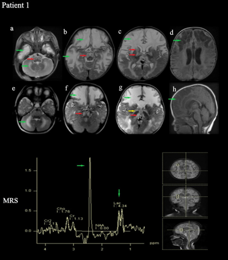
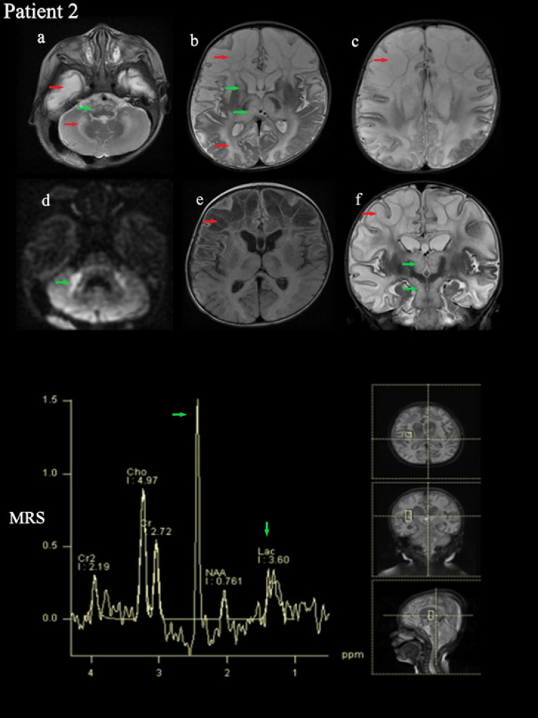
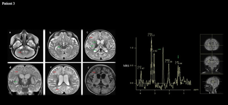
The Succinate Dehydrogenase (SDH) enzyme is known as Complex-II in the electron transport chain. This study reports the clinical and molecular investigations of three pediatric patients (two of whom are siblings), with histochemical and biochemical evidence of a severe, isolated complex II deficiency due to SDH gene mutations. The patients presented with severe hypotonia, developmental delay, spasticity, macrocephaly, and megalencephaly. Magnetic Resonance Imaging (MRI) revealed signal changes in the frontal, temporal, parietal, occipital cerebral, and cerebellar white matter, corpus striatum, thalamus, substantia nigra, inferior olivary nucleus, pyramidal tracts at the level of the pons and posterior limb of the internal capsule. Other typical findings involved a high succinate peak at 2.42 ppm and lactate peak at 1.3 ppm in Magnetic Resonance Spectroscopy (MRS). The siblings presented due to compound heterozygous c.143A>T (p. Asp48Val) and c.308T>C (p. Met103Thr) SDHB mutations, while the other patient presented due to compound heterozygous c.1754G>A (p. Arg585Gln) and c.1786G>C (p. Asp596His) SDHA mutation. The demonstration of succinate peak, particularly MRS, is highly diagnostic regarding SDH deficiency. MRS should be a standard part of routine radiological exams when there is a suspicion of a neurometabolic disease, especially mitochondrial disorders. Additionally, employing Next-Generation Sequencing (NGS) is advisable for patients as it allows for accurate diagnosis without requiring invasive procedures like muscle biopsies.

 求助内容:
求助内容: 应助结果提醒方式:


