{"title":"脊髓室管膜瘤的动态磁共振成像变化及其对手术计划的影响。","authors":"Yuki Sunohara, Yoshitaka Nagashima, Yusuke Nishimura, Masahito Hara, Hiroyuki Kato, Eisuke Tsukamoto, Kazuichi Terao, Naoto Kawaguchi, Takafumi Tanei, Ryuta Saito","doi":"10.2176/jns-nmc.2024-0328","DOIUrl":null,"url":null,"abstract":"<p><p>Spinal ependymomas are common intramedullary tumors that can show dynamic changes in magnetic resonance imaging findings over time. This study aimed to analyze these imaging changes and their implications for perioperative management. The retrospective study included patients diagnosed with World Health Organization grade 2 spinal ependymoma who underwent surgical resection and had at least 2 preoperative magnetic resonance imaging scans. Patients were divided into 2 groups based on the presence or absence of radiographic changes on magnetic resonance imaging. Magnetic resonance imaging analyses included non-contrast T1- and T2-weighted images, as well as gadolinium-enhanced T1-weighted images when available. Key features evaluated included intraparenchymal edema, hemosiderin deposition, syringomyelia, and cyst components. Changes in tumor size and contrast enhancement patterns were documented. Radiographic changes were identified in 4 out of 15 cases (26.7%). All cases with imaging changes exhibited hemosiderin deposition or hemorrhage, significantly higher than in cases without changes (100% vs. 18.2%, p < 0.05). No significant differences were observed in the presence of cystic components, syringomyelia, or edema between the groups. In the group with radiographic changes, the timeframe for these changes in the images ranged from 3 days to several years. Spinal ependymomas can demonstrate dynamic magnetic resonance imaging changes during the preoperative period, including both growth and reduction in tumor size. The presence of hemosiderin deposition or hemorrhage might be associated with these imaging changes. Proper timing of magnetic resonance imaging is crucial for informing surgical planning and optimizing treatment strategies for patients with spinal ependymomas.</p>","PeriodicalId":19225,"journal":{"name":"Neurologia medico-chirurgica","volume":" ","pages":"310-318"},"PeriodicalIF":2.3000,"publicationDate":"2025-07-15","publicationTypes":"Journal Article","fieldsOfStudy":null,"isOpenAccess":false,"openAccessPdf":"https://www.ncbi.nlm.nih.gov/pmc/articles/PMC12322308/pdf/","citationCount":"0","resultStr":"{\"title\":\"Dynamic Magnetic Resonance Imaging Changes in Spinal Ependymomas and Their Impact on Surgical Planning.\",\"authors\":\"Yuki Sunohara, Yoshitaka Nagashima, Yusuke Nishimura, Masahito Hara, Hiroyuki Kato, Eisuke Tsukamoto, Kazuichi Terao, Naoto Kawaguchi, Takafumi Tanei, Ryuta Saito\",\"doi\":\"10.2176/jns-nmc.2024-0328\",\"DOIUrl\":null,\"url\":null,\"abstract\":\"<p><p>Spinal ependymomas are common intramedullary tumors that can show dynamic changes in magnetic resonance imaging findings over time. This study aimed to analyze these imaging changes and their implications for perioperative management. The retrospective study included patients diagnosed with World Health Organization grade 2 spinal ependymoma who underwent surgical resection and had at least 2 preoperative magnetic resonance imaging scans. Patients were divided into 2 groups based on the presence or absence of radiographic changes on magnetic resonance imaging. Magnetic resonance imaging analyses included non-contrast T1- and T2-weighted images, as well as gadolinium-enhanced T1-weighted images when available. Key features evaluated included intraparenchymal edema, hemosiderin deposition, syringomyelia, and cyst components. Changes in tumor size and contrast enhancement patterns were documented. Radiographic changes were identified in 4 out of 15 cases (26.7%). All cases with imaging changes exhibited hemosiderin deposition or hemorrhage, significantly higher than in cases without changes (100% vs. 18.2%, p < 0.05). No significant differences were observed in the presence of cystic components, syringomyelia, or edema between the groups. In the group with radiographic changes, the timeframe for these changes in the images ranged from 3 days to several years. Spinal ependymomas can demonstrate dynamic magnetic resonance imaging changes during the preoperative period, including both growth and reduction in tumor size. The presence of hemosiderin deposition or hemorrhage might be associated with these imaging changes. Proper timing of magnetic resonance imaging is crucial for informing surgical planning and optimizing treatment strategies for patients with spinal ependymomas.</p>\",\"PeriodicalId\":19225,\"journal\":{\"name\":\"Neurologia medico-chirurgica\",\"volume\":\" \",\"pages\":\"310-318\"},\"PeriodicalIF\":2.3000,\"publicationDate\":\"2025-07-15\",\"publicationTypes\":\"Journal Article\",\"fieldsOfStudy\":null,\"isOpenAccess\":false,\"openAccessPdf\":\"https://www.ncbi.nlm.nih.gov/pmc/articles/PMC12322308/pdf/\",\"citationCount\":\"0\",\"resultStr\":null,\"platform\":\"Semanticscholar\",\"paperid\":null,\"PeriodicalName\":\"Neurologia medico-chirurgica\",\"FirstCategoryId\":\"3\",\"ListUrlMain\":\"https://doi.org/10.2176/jns-nmc.2024-0328\",\"RegionNum\":4,\"RegionCategory\":\"医学\",\"ArticlePicture\":[],\"TitleCN\":null,\"AbstractTextCN\":null,\"PMCID\":null,\"EPubDate\":\"2025/5/29 0:00:00\",\"PubModel\":\"Epub\",\"JCR\":\"Q2\",\"JCRName\":\"CLINICAL NEUROLOGY\",\"Score\":null,\"Total\":0}","platform":"Semanticscholar","paperid":null,"PeriodicalName":"Neurologia medico-chirurgica","FirstCategoryId":"3","ListUrlMain":"https://doi.org/10.2176/jns-nmc.2024-0328","RegionNum":4,"RegionCategory":"医学","ArticlePicture":[],"TitleCN":null,"AbstractTextCN":null,"PMCID":null,"EPubDate":"2025/5/29 0:00:00","PubModel":"Epub","JCR":"Q2","JCRName":"CLINICAL NEUROLOGY","Score":null,"Total":0}
引用次数: 0
摘要
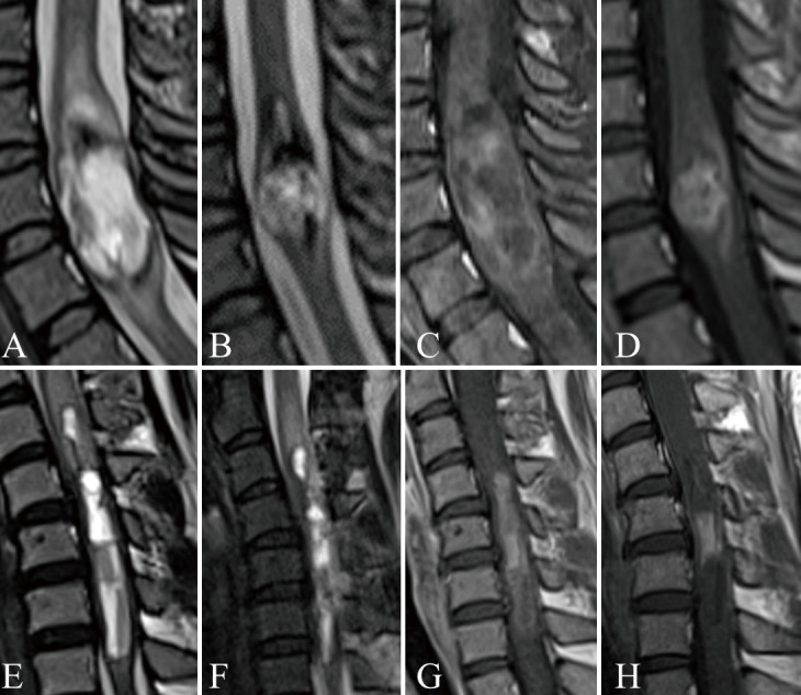
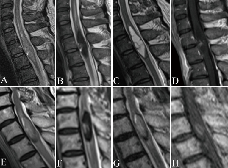
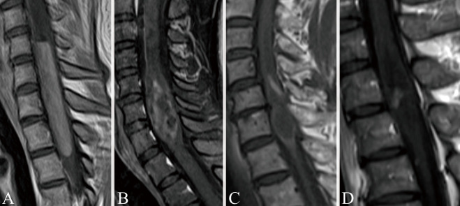
脊髓室管膜瘤是常见的髓内肿瘤,其磁共振成像表现随时间的变化而变化。本研究旨在分析这些影像学变化及其对围手术期处理的意义。该回顾性研究纳入了世界卫生组织诊断为2级脊髓室管膜瘤的患者,这些患者接受了手术切除,术前至少进行了2次磁共振成像扫描。根据磁共振成像有无影像学改变将患者分为两组。磁共振成像分析包括非对比T1和t2加权图像,以及可用的钆增强T1加权图像。评估的主要特征包括实质内水肿、含铁血黄素沉积、脊髓空洞和囊肿成分。肿瘤大小的变化和对比增强模式被记录下来。15例患者中有4例(26.7%)有影像学改变。所有有影像学改变的患者均表现为含铁血黄素沉积或出血,明显高于无影像学改变的患者(100% vs. 18.2%, p < 0.05)。在囊性成分、脊髓空洞或水肿方面,两组之间没有显著差异。在有影像学改变的组中,这些图像变化的时间范围从3天到几年不等。脊髓室管膜瘤可以在术前表现出动态的磁共振成像变化,包括肿瘤大小的生长和缩小。含铁血黄素沉积或出血的存在可能与这些影像学改变有关。适当的时间磁共振成像是至关重要的,告知手术计划和优化治疗策略的患者的脊髓室管膜瘤。
Dynamic Magnetic Resonance Imaging Changes in Spinal Ependymomas and Their Impact on Surgical Planning.
Spinal ependymomas are common intramedullary tumors that can show dynamic changes in magnetic resonance imaging findings over time. This study aimed to analyze these imaging changes and their implications for perioperative management. The retrospective study included patients diagnosed with World Health Organization grade 2 spinal ependymoma who underwent surgical resection and had at least 2 preoperative magnetic resonance imaging scans. Patients were divided into 2 groups based on the presence or absence of radiographic changes on magnetic resonance imaging. Magnetic resonance imaging analyses included non-contrast T1- and T2-weighted images, as well as gadolinium-enhanced T1-weighted images when available. Key features evaluated included intraparenchymal edema, hemosiderin deposition, syringomyelia, and cyst components. Changes in tumor size and contrast enhancement patterns were documented. Radiographic changes were identified in 4 out of 15 cases (26.7%). All cases with imaging changes exhibited hemosiderin deposition or hemorrhage, significantly higher than in cases without changes (100% vs. 18.2%, p < 0.05). No significant differences were observed in the presence of cystic components, syringomyelia, or edema between the groups. In the group with radiographic changes, the timeframe for these changes in the images ranged from 3 days to several years. Spinal ependymomas can demonstrate dynamic magnetic resonance imaging changes during the preoperative period, including both growth and reduction in tumor size. The presence of hemosiderin deposition or hemorrhage might be associated with these imaging changes. Proper timing of magnetic resonance imaging is crucial for informing surgical planning and optimizing treatment strategies for patients with spinal ependymomas.
 求助内容:
求助内容: 应助结果提醒方式:


