Alejandra Avila, Varsha Thakur, Natalie Vincent, Pilar Valencia, Mecker G Möller, Rimpi Khurana, Guo Yan, Jennifer C Tang, Barbara Bedogni, Natalia Jaimes
{"title":"长期晒伤皮肤上的黑色素瘤:解读基因表达特征。","authors":"Alejandra Avila, Varsha Thakur, Natalie Vincent, Pilar Valencia, Mecker G Möller, Rimpi Khurana, Guo Yan, Jennifer C Tang, Barbara Bedogni, Natalia Jaimes","doi":"10.5826/dpc.1502a4952","DOIUrl":null,"url":null,"abstract":"<p><strong>Introduction: </strong>Melanoma of the skin is responsible for most skin cancer-related deaths. It is well known that exposure to ultraviolet radiation is the most common and modifiable risk factor for melanoma. Melanomas arising on chronically sun-damaged skin (CSDS) have shown a higher mutational burden.</p><p><strong>Objectives: </strong>To analyze skin samples of patients with melanoma on CSDS to identify possible gene expression signatures that may contribute to melanomagenesis.</p><p><strong>Methods: </strong>This experimental observational analysis, conducted at the Dermatology Melanoma and Pigmented Lesion Clinic at University of Miami Hospitals/Sylvester Comprehensive Cancer Center, Miami, Florida, included a total of 10 patients over 18 years of age with a recent diagnosis of melanoma on CSDS. For each patient, two skin samples were obtained using a 2-mm punch (one from CSDS within 2 cm of the primary melanoma, another from sun-protected skin). Skin samples were sent to the Sylvester Onco-genomics Shared Resource (OGSR) for library preparation and RNA sequencing. Main outcome was the identification of differentially expressed genes between CSDS and non-CSDS of patients with a recent diagnosis of melanoma.</p><p><strong>Results: </strong>A total of four skin samples met the necessary quality standards for molecular analyses. Significant differences were observed between the CSDS and non-CSDS samples. Pathways involved in inflammation (e.g., IL-17 signaling), immune responses (e.g., ABC transporters), and oxidative phosphorylation were overexpressed in CSDS.</p><p><strong>Conclusions: </strong>CSDS can be an adequate milieu for the development and progression of melanoma. CSDS reveals overexpression of pathways involved in inflammation, immune responses, and oxidative phosphorylation, all of which may facilitate interactions between the skin microenvironment and melanocytes/melanoma cells, predisposing to melanoma development and progression.</p>","PeriodicalId":11168,"journal":{"name":"Dermatology practical & conceptual","volume":"15 2","pages":""},"PeriodicalIF":2.3000,"publicationDate":"2025-04-01","publicationTypes":"Journal Article","fieldsOfStudy":null,"isOpenAccess":false,"openAccessPdf":"https://www.ncbi.nlm.nih.gov/pmc/articles/PMC12090909/pdf/","citationCount":"0","resultStr":"{\"title\":\"Melanoma on Chronically Sun-Damaged Skin: Deciphering Gene Expression Signatures.\",\"authors\":\"Alejandra Avila, Varsha Thakur, Natalie Vincent, Pilar Valencia, Mecker G Möller, Rimpi Khurana, Guo Yan, Jennifer C Tang, Barbara Bedogni, Natalia Jaimes\",\"doi\":\"10.5826/dpc.1502a4952\",\"DOIUrl\":null,\"url\":null,\"abstract\":\"<p><strong>Introduction: </strong>Melanoma of the skin is responsible for most skin cancer-related deaths. It is well known that exposure to ultraviolet radiation is the most common and modifiable risk factor for melanoma. Melanomas arising on chronically sun-damaged skin (CSDS) have shown a higher mutational burden.</p><p><strong>Objectives: </strong>To analyze skin samples of patients with melanoma on CSDS to identify possible gene expression signatures that may contribute to melanomagenesis.</p><p><strong>Methods: </strong>This experimental observational analysis, conducted at the Dermatology Melanoma and Pigmented Lesion Clinic at University of Miami Hospitals/Sylvester Comprehensive Cancer Center, Miami, Florida, included a total of 10 patients over 18 years of age with a recent diagnosis of melanoma on CSDS. For each patient, two skin samples were obtained using a 2-mm punch (one from CSDS within 2 cm of the primary melanoma, another from sun-protected skin). Skin samples were sent to the Sylvester Onco-genomics Shared Resource (OGSR) for library preparation and RNA sequencing. Main outcome was the identification of differentially expressed genes between CSDS and non-CSDS of patients with a recent diagnosis of melanoma.</p><p><strong>Results: </strong>A total of four skin samples met the necessary quality standards for molecular analyses. Significant differences were observed between the CSDS and non-CSDS samples. Pathways involved in inflammation (e.g., IL-17 signaling), immune responses (e.g., ABC transporters), and oxidative phosphorylation were overexpressed in CSDS.</p><p><strong>Conclusions: </strong>CSDS can be an adequate milieu for the development and progression of melanoma. CSDS reveals overexpression of pathways involved in inflammation, immune responses, and oxidative phosphorylation, all of which may facilitate interactions between the skin microenvironment and melanocytes/melanoma cells, predisposing to melanoma development and progression.</p>\",\"PeriodicalId\":11168,\"journal\":{\"name\":\"Dermatology practical & conceptual\",\"volume\":\"15 2\",\"pages\":\"\"},\"PeriodicalIF\":2.3000,\"publicationDate\":\"2025-04-01\",\"publicationTypes\":\"Journal Article\",\"fieldsOfStudy\":null,\"isOpenAccess\":false,\"openAccessPdf\":\"https://www.ncbi.nlm.nih.gov/pmc/articles/PMC12090909/pdf/\",\"citationCount\":\"0\",\"resultStr\":null,\"platform\":\"Semanticscholar\",\"paperid\":null,\"PeriodicalName\":\"Dermatology practical & conceptual\",\"FirstCategoryId\":\"3\",\"ListUrlMain\":\"https://doi.org/10.5826/dpc.1502a4952\",\"RegionNum\":4,\"RegionCategory\":\"医学\",\"ArticlePicture\":[],\"TitleCN\":null,\"AbstractTextCN\":null,\"PMCID\":null,\"EPubDate\":\"\",\"PubModel\":\"\",\"JCR\":\"Q2\",\"JCRName\":\"DERMATOLOGY\",\"Score\":null,\"Total\":0}","platform":"Semanticscholar","paperid":null,"PeriodicalName":"Dermatology practical & conceptual","FirstCategoryId":"3","ListUrlMain":"https://doi.org/10.5826/dpc.1502a4952","RegionNum":4,"RegionCategory":"医学","ArticlePicture":[],"TitleCN":null,"AbstractTextCN":null,"PMCID":null,"EPubDate":"","PubModel":"","JCR":"Q2","JCRName":"DERMATOLOGY","Score":null,"Total":0}
Melanoma on Chronically Sun-Damaged Skin: Deciphering Gene Expression Signatures.
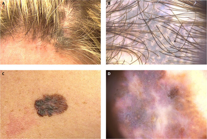
Introduction: Melanoma of the skin is responsible for most skin cancer-related deaths. It is well known that exposure to ultraviolet radiation is the most common and modifiable risk factor for melanoma. Melanomas arising on chronically sun-damaged skin (CSDS) have shown a higher mutational burden.
Objectives: To analyze skin samples of patients with melanoma on CSDS to identify possible gene expression signatures that may contribute to melanomagenesis.
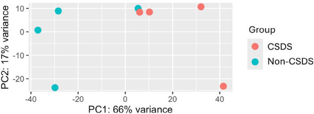
Methods: This experimental observational analysis, conducted at the Dermatology Melanoma and Pigmented Lesion Clinic at University of Miami Hospitals/Sylvester Comprehensive Cancer Center, Miami, Florida, included a total of 10 patients over 18 years of age with a recent diagnosis of melanoma on CSDS. For each patient, two skin samples were obtained using a 2-mm punch (one from CSDS within 2 cm of the primary melanoma, another from sun-protected skin). Skin samples were sent to the Sylvester Onco-genomics Shared Resource (OGSR) for library preparation and RNA sequencing. Main outcome was the identification of differentially expressed genes between CSDS and non-CSDS of patients with a recent diagnosis of melanoma.
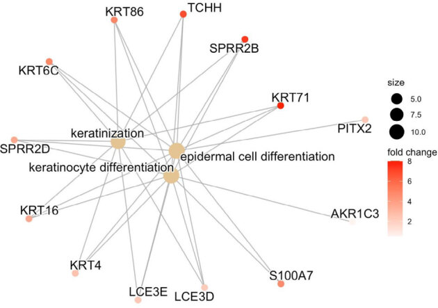
Results: A total of four skin samples met the necessary quality standards for molecular analyses. Significant differences were observed between the CSDS and non-CSDS samples. Pathways involved in inflammation (e.g., IL-17 signaling), immune responses (e.g., ABC transporters), and oxidative phosphorylation were overexpressed in CSDS.
Conclusions: CSDS can be an adequate milieu for the development and progression of melanoma. CSDS reveals overexpression of pathways involved in inflammation, immune responses, and oxidative phosphorylation, all of which may facilitate interactions between the skin microenvironment and melanocytes/melanoma cells, predisposing to melanoma development and progression.
 求助内容:
求助内容: 应助结果提醒方式:


