Camila Scharf, Aleksandra A Stefaniak, Mario Vaccaro, Francesco Borgia, Federico Vaccaro, Fabrizio Chirico, Andrea Battisti, Valentino Valentini, Giuseppe Argenziano
{"title":"模拟鳞状细胞癌的头皮糜烂性脓疱性皮肤病:皮肤镜检查有帮助吗?","authors":"Camila Scharf, Aleksandra A Stefaniak, Mario Vaccaro, Francesco Borgia, Federico Vaccaro, Fabrizio Chirico, Andrea Battisti, Valentino Valentini, Giuseppe Argenziano","doi":"10.5826/dpc.1502a4796","DOIUrl":null,"url":null,"abstract":"<p><strong>Introduction: </strong>Erosive pustular dermatosis of the scalp (EPDS) presents as a rare and challenging skin disorder marked by erosions, pustules, and crusted lesions on the scalp. Diagnosing EPDS is complex, often mimicking other dermatological conditions like actinic keratosis (AK), basal cell carcinoma (BCC), or squamous cell carcinoma (SCC). Therapeutic challenges arise from EPDS's chronic, relapsing nature, which requires long-term management.</p><p><strong>Objective: </strong>This study aimed to identify and compare dermoscopic features of EPDS mimicking SCC of the scalp to improve diagnostic accuracy and support non-invasive differentiation.</p><p><strong>Methods: </strong>A retrospective descriptive study, conducted in two Italian dermatological centers from 2017 to 2023, included 43 patients initially diagnosed with SCC of the scalp that was afterwards confirmed as EPDS through either histology or clinical evaluation and follow-up. Clinical and dermoscopic criteria were evaluated. A comparison of dermoscopical criteria with the same number of confirmed SCC of the scalp was performed.</p><p><strong>Results: </strong>A total of 43 patients were included, predominantly male (42:1). Androgenetic alopecia was present in 65% of cases, predominantly in the parietal area (44%). Hairpin and dotted vessels, along with white and combined lesion colors, were strongly associated with SCC, while polymorphic and branched vessels, orange tones, and targetoid hair follicles predominated in EPDS.</p><p><strong>Conclusions: </strong>EPDS poses diagnostic and therapeutic challenges due to its unique features and elusive etiology. Distinguishing SCC from EPDS is crucial to avoid unnecessary treatments; dermoscopy can serve as an instrument in this process.</p>","PeriodicalId":11168,"journal":{"name":"Dermatology practical & conceptual","volume":"15 2","pages":""},"PeriodicalIF":2.3000,"publicationDate":"2025-04-01","publicationTypes":"Journal Article","fieldsOfStudy":null,"isOpenAccess":false,"openAccessPdf":"https://www.ncbi.nlm.nih.gov/pmc/articles/PMC12090949/pdf/","citationCount":"0","resultStr":"{\"title\":\"Erosive Pustular Dermatosis of the Scalp Mimicking Squamous Cell Carcinoma: Can Dermoscopy Be Helpful?\",\"authors\":\"Camila Scharf, Aleksandra A Stefaniak, Mario Vaccaro, Francesco Borgia, Federico Vaccaro, Fabrizio Chirico, Andrea Battisti, Valentino Valentini, Giuseppe Argenziano\",\"doi\":\"10.5826/dpc.1502a4796\",\"DOIUrl\":null,\"url\":null,\"abstract\":\"<p><strong>Introduction: </strong>Erosive pustular dermatosis of the scalp (EPDS) presents as a rare and challenging skin disorder marked by erosions, pustules, and crusted lesions on the scalp. Diagnosing EPDS is complex, often mimicking other dermatological conditions like actinic keratosis (AK), basal cell carcinoma (BCC), or squamous cell carcinoma (SCC). Therapeutic challenges arise from EPDS's chronic, relapsing nature, which requires long-term management.</p><p><strong>Objective: </strong>This study aimed to identify and compare dermoscopic features of EPDS mimicking SCC of the scalp to improve diagnostic accuracy and support non-invasive differentiation.</p><p><strong>Methods: </strong>A retrospective descriptive study, conducted in two Italian dermatological centers from 2017 to 2023, included 43 patients initially diagnosed with SCC of the scalp that was afterwards confirmed as EPDS through either histology or clinical evaluation and follow-up. Clinical and dermoscopic criteria were evaluated. A comparison of dermoscopical criteria with the same number of confirmed SCC of the scalp was performed.</p><p><strong>Results: </strong>A total of 43 patients were included, predominantly male (42:1). Androgenetic alopecia was present in 65% of cases, predominantly in the parietal area (44%). Hairpin and dotted vessels, along with white and combined lesion colors, were strongly associated with SCC, while polymorphic and branched vessels, orange tones, and targetoid hair follicles predominated in EPDS.</p><p><strong>Conclusions: </strong>EPDS poses diagnostic and therapeutic challenges due to its unique features and elusive etiology. Distinguishing SCC from EPDS is crucial to avoid unnecessary treatments; dermoscopy can serve as an instrument in this process.</p>\",\"PeriodicalId\":11168,\"journal\":{\"name\":\"Dermatology practical & conceptual\",\"volume\":\"15 2\",\"pages\":\"\"},\"PeriodicalIF\":2.3000,\"publicationDate\":\"2025-04-01\",\"publicationTypes\":\"Journal Article\",\"fieldsOfStudy\":null,\"isOpenAccess\":false,\"openAccessPdf\":\"https://www.ncbi.nlm.nih.gov/pmc/articles/PMC12090949/pdf/\",\"citationCount\":\"0\",\"resultStr\":null,\"platform\":\"Semanticscholar\",\"paperid\":null,\"PeriodicalName\":\"Dermatology practical & conceptual\",\"FirstCategoryId\":\"3\",\"ListUrlMain\":\"https://doi.org/10.5826/dpc.1502a4796\",\"RegionNum\":4,\"RegionCategory\":\"医学\",\"ArticlePicture\":[],\"TitleCN\":null,\"AbstractTextCN\":null,\"PMCID\":null,\"EPubDate\":\"\",\"PubModel\":\"\",\"JCR\":\"Q2\",\"JCRName\":\"DERMATOLOGY\",\"Score\":null,\"Total\":0}","platform":"Semanticscholar","paperid":null,"PeriodicalName":"Dermatology practical & conceptual","FirstCategoryId":"3","ListUrlMain":"https://doi.org/10.5826/dpc.1502a4796","RegionNum":4,"RegionCategory":"医学","ArticlePicture":[],"TitleCN":null,"AbstractTextCN":null,"PMCID":null,"EPubDate":"","PubModel":"","JCR":"Q2","JCRName":"DERMATOLOGY","Score":null,"Total":0}
Erosive Pustular Dermatosis of the Scalp Mimicking Squamous Cell Carcinoma: Can Dermoscopy Be Helpful?
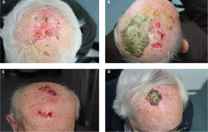
Introduction: Erosive pustular dermatosis of the scalp (EPDS) presents as a rare and challenging skin disorder marked by erosions, pustules, and crusted lesions on the scalp. Diagnosing EPDS is complex, often mimicking other dermatological conditions like actinic keratosis (AK), basal cell carcinoma (BCC), or squamous cell carcinoma (SCC). Therapeutic challenges arise from EPDS's chronic, relapsing nature, which requires long-term management.
Objective: This study aimed to identify and compare dermoscopic features of EPDS mimicking SCC of the scalp to improve diagnostic accuracy and support non-invasive differentiation.
Methods: A retrospective descriptive study, conducted in two Italian dermatological centers from 2017 to 2023, included 43 patients initially diagnosed with SCC of the scalp that was afterwards confirmed as EPDS through either histology or clinical evaluation and follow-up. Clinical and dermoscopic criteria were evaluated. A comparison of dermoscopical criteria with the same number of confirmed SCC of the scalp was performed.
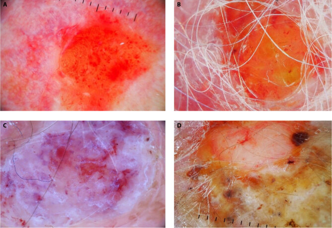
Results: A total of 43 patients were included, predominantly male (42:1). Androgenetic alopecia was present in 65% of cases, predominantly in the parietal area (44%). Hairpin and dotted vessels, along with white and combined lesion colors, were strongly associated with SCC, while polymorphic and branched vessels, orange tones, and targetoid hair follicles predominated in EPDS.
Conclusions: EPDS poses diagnostic and therapeutic challenges due to its unique features and elusive etiology. Distinguishing SCC from EPDS is crucial to avoid unnecessary treatments; dermoscopy can serve as an instrument in this process.

 求助内容:
求助内容: 应助结果提醒方式:


