Oluwatodimu Richard Raji, Jason E Pope, Steven M Falowski, Michael Stoffman, Jeremi M Leasure
{"title":"骶髂关节的固定:基于尸体的后路插入和后外侧经骨技术的同步控制生物力学比较。","authors":"Oluwatodimu Richard Raji, Jason E Pope, Steven M Falowski, Michael Stoffman, Jeremi M Leasure","doi":"10.14245/ns.2448940.470","DOIUrl":null,"url":null,"abstract":"<p><strong>Objective: </strong>Our study aimed to compare the posterior interposition technique against the posterolateral transosseous technique in the same cadaver specimens.</p><p><strong>Methods: </strong>Computer and cadaver models of 2 fixation techniques were developed. The computer model was constructed to analyze bone volume removed during implant placement and the bony surface area available for fusion. The cadaver model included quasi-static multidirectional bending flexibility and dynamic fatigue loading. Relative motions between the sacrum and ilium were measured intact, after joint destabilization, after fixation with direct-posterior and posterolateral techniques, and after 18,500 cycles of fatigue loading. Relative positions between each implant and the sacrum and ilium were measured after fixation and fatigue loading to ascertain the quality of the bone-implant interface. The 2 techniques were randomized to the left and right sacroiliac joints of the same cadavers.</p><p><strong>Results: </strong>The posterior interposition technique removed less bone volume and facilitated a larger surface area available for bony fusion. Posterior interposition significantly reduced the nutation/counternutation motion of the sacroiliac joint (42% ± 8%) and reduced it more than the posterolateral transosseous technique (14% ± 4%). Upon fatigue loading, the posterior interposition implant maintained the bone-implant interface across all specimens, while the posterolateral transosseous implant migrated or subsided in 20%-50% of specimens.</p><p><strong>Conclusion: </strong>Posterior interposition fixation of the sacroiliac joint reduces joint motion. The amount of fixation from the posterior technique is superior and more durable than the amount of fixation achieved by the posterolateral technique.</p>","PeriodicalId":19269,"journal":{"name":"Neurospine","volume":"22 1","pages":"185-193"},"PeriodicalIF":3.6000,"publicationDate":"2025-03-01","publicationTypes":"Journal Article","fieldsOfStudy":null,"isOpenAccess":false,"openAccessPdf":"https://www.ncbi.nlm.nih.gov/pmc/articles/PMC12010861/pdf/","citationCount":"0","resultStr":"{\"title\":\"Fixation of the Sacroiliac Joint: A Cadaver-Based Concurrent-Controlled Biomechanical Comparison of Posterior Interposition and Posterolateral Transosseous Techniques.\",\"authors\":\"Oluwatodimu Richard Raji, Jason E Pope, Steven M Falowski, Michael Stoffman, Jeremi M Leasure\",\"doi\":\"10.14245/ns.2448940.470\",\"DOIUrl\":null,\"url\":null,\"abstract\":\"<p><strong>Objective: </strong>Our study aimed to compare the posterior interposition technique against the posterolateral transosseous technique in the same cadaver specimens.</p><p><strong>Methods: </strong>Computer and cadaver models of 2 fixation techniques were developed. The computer model was constructed to analyze bone volume removed during implant placement and the bony surface area available for fusion. The cadaver model included quasi-static multidirectional bending flexibility and dynamic fatigue loading. Relative motions between the sacrum and ilium were measured intact, after joint destabilization, after fixation with direct-posterior and posterolateral techniques, and after 18,500 cycles of fatigue loading. Relative positions between each implant and the sacrum and ilium were measured after fixation and fatigue loading to ascertain the quality of the bone-implant interface. The 2 techniques were randomized to the left and right sacroiliac joints of the same cadavers.</p><p><strong>Results: </strong>The posterior interposition technique removed less bone volume and facilitated a larger surface area available for bony fusion. Posterior interposition significantly reduced the nutation/counternutation motion of the sacroiliac joint (42% ± 8%) and reduced it more than the posterolateral transosseous technique (14% ± 4%). Upon fatigue loading, the posterior interposition implant maintained the bone-implant interface across all specimens, while the posterolateral transosseous implant migrated or subsided in 20%-50% of specimens.</p><p><strong>Conclusion: </strong>Posterior interposition fixation of the sacroiliac joint reduces joint motion. The amount of fixation from the posterior technique is superior and more durable than the amount of fixation achieved by the posterolateral technique.</p>\",\"PeriodicalId\":19269,\"journal\":{\"name\":\"Neurospine\",\"volume\":\"22 1\",\"pages\":\"185-193\"},\"PeriodicalIF\":3.6000,\"publicationDate\":\"2025-03-01\",\"publicationTypes\":\"Journal Article\",\"fieldsOfStudy\":null,\"isOpenAccess\":false,\"openAccessPdf\":\"https://www.ncbi.nlm.nih.gov/pmc/articles/PMC12010861/pdf/\",\"citationCount\":\"0\",\"resultStr\":null,\"platform\":\"Semanticscholar\",\"paperid\":null,\"PeriodicalName\":\"Neurospine\",\"FirstCategoryId\":\"3\",\"ListUrlMain\":\"https://doi.org/10.14245/ns.2448940.470\",\"RegionNum\":2,\"RegionCategory\":\"医学\",\"ArticlePicture\":[],\"TitleCN\":null,\"AbstractTextCN\":null,\"PMCID\":null,\"EPubDate\":\"2025/3/31 0:00:00\",\"PubModel\":\"Epub\",\"JCR\":\"Q1\",\"JCRName\":\"CLINICAL NEUROLOGY\",\"Score\":null,\"Total\":0}","platform":"Semanticscholar","paperid":null,"PeriodicalName":"Neurospine","FirstCategoryId":"3","ListUrlMain":"https://doi.org/10.14245/ns.2448940.470","RegionNum":2,"RegionCategory":"医学","ArticlePicture":[],"TitleCN":null,"AbstractTextCN":null,"PMCID":null,"EPubDate":"2025/3/31 0:00:00","PubModel":"Epub","JCR":"Q1","JCRName":"CLINICAL NEUROLOGY","Score":null,"Total":0}
Fixation of the Sacroiliac Joint: A Cadaver-Based Concurrent-Controlled Biomechanical Comparison of Posterior Interposition and Posterolateral Transosseous Techniques.
Objective: Our study aimed to compare the posterior interposition technique against the posterolateral transosseous technique in the same cadaver specimens.
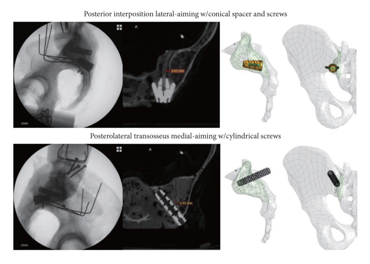
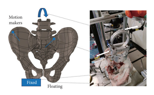
Methods: Computer and cadaver models of 2 fixation techniques were developed. The computer model was constructed to analyze bone volume removed during implant placement and the bony surface area available for fusion. The cadaver model included quasi-static multidirectional bending flexibility and dynamic fatigue loading. Relative motions between the sacrum and ilium were measured intact, after joint destabilization, after fixation with direct-posterior and posterolateral techniques, and after 18,500 cycles of fatigue loading. Relative positions between each implant and the sacrum and ilium were measured after fixation and fatigue loading to ascertain the quality of the bone-implant interface. The 2 techniques were randomized to the left and right sacroiliac joints of the same cadavers.
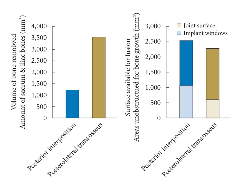
Results: The posterior interposition technique removed less bone volume and facilitated a larger surface area available for bony fusion. Posterior interposition significantly reduced the nutation/counternutation motion of the sacroiliac joint (42% ± 8%) and reduced it more than the posterolateral transosseous technique (14% ± 4%). Upon fatigue loading, the posterior interposition implant maintained the bone-implant interface across all specimens, while the posterolateral transosseous implant migrated or subsided in 20%-50% of specimens.
Conclusion: Posterior interposition fixation of the sacroiliac joint reduces joint motion. The amount of fixation from the posterior technique is superior and more durable than the amount of fixation achieved by the posterolateral technique.

 求助内容:
求助内容: 应助结果提醒方式:


