Siyuan Qin, Ruomu Qu, Ke Liu, Ruixin Yan, Weili Zhao, Jun Xu, Enlong Zhang, Feifei Zhou, Ning Lang
{"title":"使用可解释的放射组学模型预测颈椎后纵韧带骨化的术后进展。","authors":"Siyuan Qin, Ruomu Qu, Ke Liu, Ruixin Yan, Weili Zhao, Jun Xu, Enlong Zhang, Feifei Zhou, Ning Lang","doi":"10.14245/ns.2448846.423","DOIUrl":null,"url":null,"abstract":"<p><strong>Objective: </strong>This study investigates the potential of radiomics to predict postoperative progression of ossification of the posterior longitudinal ligament (OPLL) after posterior cervical spine surgery.</p><p><strong>Methods: </strong>This retrospective study included 473 patients diagnosed with OPLL at Peking University Third Hospital between October 2006 and September 2022. Patients underwent posterior spinal surgery and had at least 2 computed tomography (CT) examinations spaced at least 1 year apart. OPLL progression was defined as an annual growth rate exceeding 7.5%. Radiomic features were extracted from preoperative CT images of the OPLL lesions, followed by feature selection using correlation coefficient analysis and least absolute shrinkage and selection operator, and dimensionality reduction using principal component analysis. Univariable analysis identified significant clinical variables for constructing the clinical model. Logistic regression models, including the Rad-score model, clinical model, and combined model, were developed to predict OPLL progression.</p><p><strong>Results: </strong>Of the 473 patients, 191 (40.4%) experienced OPLL progression. On the testing set, the combined model, which incorporated the Rad-score and clinical variables (area under the receiver operating characteristic curve [AUC] = 0.751), outperformed both the radiomics-only model (AUC = 0.693) and the clinical model (AUC = 0.620). Calibration curves demonstrated good agreement between predicted probabilities and observed outcomes, and decision curve analysis confirmed the clinical utility of the combined model. SHAP (SHapley Additive exPlanations) analysis indicated that the Rad-score and age were key contributors to the model's predictions, enhancing clinical interpretability.</p><p><strong>Conclusion: </strong>Radiomics, combined with clinical variables, provides a valuable predictive tool for assessing the risk of postoperative progression in cervical OPLL, supporting more personalized treatment strategies. Prospective, multicenter validation is needed to confirm the utility of the model in broader clinical settings.</p>","PeriodicalId":19269,"journal":{"name":"Neurospine","volume":"22 1","pages":"144-156"},"PeriodicalIF":3.6000,"publicationDate":"2025-03-01","publicationTypes":"Journal Article","fieldsOfStudy":null,"isOpenAccess":false,"openAccessPdf":"https://www.ncbi.nlm.nih.gov/pmc/articles/PMC12010848/pdf/","citationCount":"0","resultStr":"{\"title\":\"Predicting Postoperative Progression of Ossification of the Posterior Longitudinal Ligament in the Cervical Spine Using Interpretable Radiomics Models.\",\"authors\":\"Siyuan Qin, Ruomu Qu, Ke Liu, Ruixin Yan, Weili Zhao, Jun Xu, Enlong Zhang, Feifei Zhou, Ning Lang\",\"doi\":\"10.14245/ns.2448846.423\",\"DOIUrl\":null,\"url\":null,\"abstract\":\"<p><strong>Objective: </strong>This study investigates the potential of radiomics to predict postoperative progression of ossification of the posterior longitudinal ligament (OPLL) after posterior cervical spine surgery.</p><p><strong>Methods: </strong>This retrospective study included 473 patients diagnosed with OPLL at Peking University Third Hospital between October 2006 and September 2022. Patients underwent posterior spinal surgery and had at least 2 computed tomography (CT) examinations spaced at least 1 year apart. OPLL progression was defined as an annual growth rate exceeding 7.5%. Radiomic features were extracted from preoperative CT images of the OPLL lesions, followed by feature selection using correlation coefficient analysis and least absolute shrinkage and selection operator, and dimensionality reduction using principal component analysis. Univariable analysis identified significant clinical variables for constructing the clinical model. Logistic regression models, including the Rad-score model, clinical model, and combined model, were developed to predict OPLL progression.</p><p><strong>Results: </strong>Of the 473 patients, 191 (40.4%) experienced OPLL progression. On the testing set, the combined model, which incorporated the Rad-score and clinical variables (area under the receiver operating characteristic curve [AUC] = 0.751), outperformed both the radiomics-only model (AUC = 0.693) and the clinical model (AUC = 0.620). Calibration curves demonstrated good agreement between predicted probabilities and observed outcomes, and decision curve analysis confirmed the clinical utility of the combined model. SHAP (SHapley Additive exPlanations) analysis indicated that the Rad-score and age were key contributors to the model's predictions, enhancing clinical interpretability.</p><p><strong>Conclusion: </strong>Radiomics, combined with clinical variables, provides a valuable predictive tool for assessing the risk of postoperative progression in cervical OPLL, supporting more personalized treatment strategies. Prospective, multicenter validation is needed to confirm the utility of the model in broader clinical settings.</p>\",\"PeriodicalId\":19269,\"journal\":{\"name\":\"Neurospine\",\"volume\":\"22 1\",\"pages\":\"144-156\"},\"PeriodicalIF\":3.6000,\"publicationDate\":\"2025-03-01\",\"publicationTypes\":\"Journal Article\",\"fieldsOfStudy\":null,\"isOpenAccess\":false,\"openAccessPdf\":\"https://www.ncbi.nlm.nih.gov/pmc/articles/PMC12010848/pdf/\",\"citationCount\":\"0\",\"resultStr\":null,\"platform\":\"Semanticscholar\",\"paperid\":null,\"PeriodicalName\":\"Neurospine\",\"FirstCategoryId\":\"3\",\"ListUrlMain\":\"https://doi.org/10.14245/ns.2448846.423\",\"RegionNum\":2,\"RegionCategory\":\"医学\",\"ArticlePicture\":[],\"TitleCN\":null,\"AbstractTextCN\":null,\"PMCID\":null,\"EPubDate\":\"2025/3/31 0:00:00\",\"PubModel\":\"Epub\",\"JCR\":\"Q1\",\"JCRName\":\"CLINICAL NEUROLOGY\",\"Score\":null,\"Total\":0}","platform":"Semanticscholar","paperid":null,"PeriodicalName":"Neurospine","FirstCategoryId":"3","ListUrlMain":"https://doi.org/10.14245/ns.2448846.423","RegionNum":2,"RegionCategory":"医学","ArticlePicture":[],"TitleCN":null,"AbstractTextCN":null,"PMCID":null,"EPubDate":"2025/3/31 0:00:00","PubModel":"Epub","JCR":"Q1","JCRName":"CLINICAL NEUROLOGY","Score":null,"Total":0}
Predicting Postoperative Progression of Ossification of the Posterior Longitudinal Ligament in the Cervical Spine Using Interpretable Radiomics Models.
Objective: This study investigates the potential of radiomics to predict postoperative progression of ossification of the posterior longitudinal ligament (OPLL) after posterior cervical spine surgery.
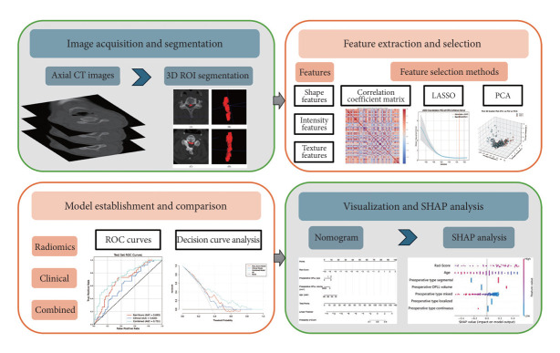
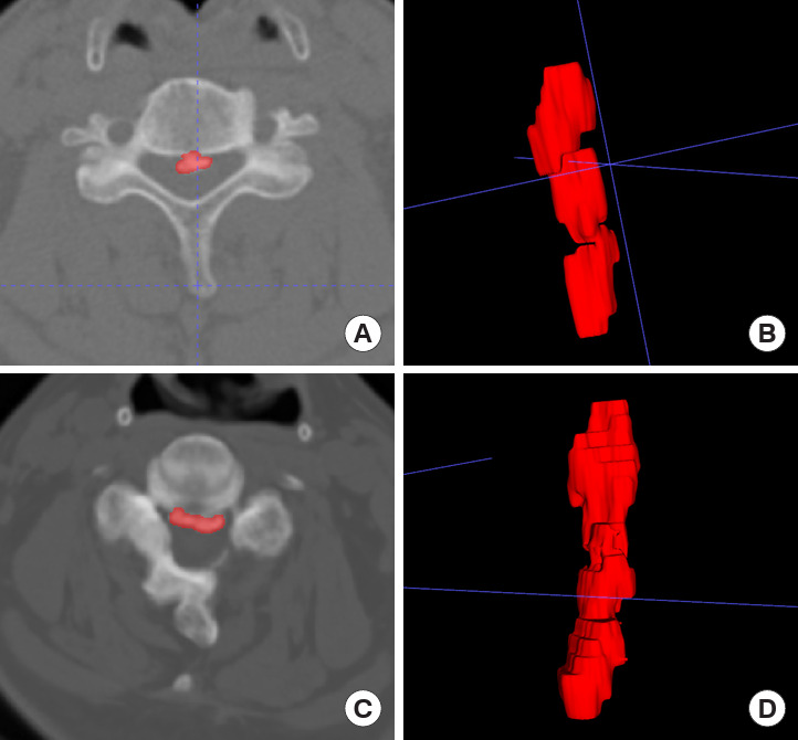
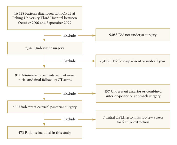
Methods: This retrospective study included 473 patients diagnosed with OPLL at Peking University Third Hospital between October 2006 and September 2022. Patients underwent posterior spinal surgery and had at least 2 computed tomography (CT) examinations spaced at least 1 year apart. OPLL progression was defined as an annual growth rate exceeding 7.5%. Radiomic features were extracted from preoperative CT images of the OPLL lesions, followed by feature selection using correlation coefficient analysis and least absolute shrinkage and selection operator, and dimensionality reduction using principal component analysis. Univariable analysis identified significant clinical variables for constructing the clinical model. Logistic regression models, including the Rad-score model, clinical model, and combined model, were developed to predict OPLL progression.
Results: Of the 473 patients, 191 (40.4%) experienced OPLL progression. On the testing set, the combined model, which incorporated the Rad-score and clinical variables (area under the receiver operating characteristic curve [AUC] = 0.751), outperformed both the radiomics-only model (AUC = 0.693) and the clinical model (AUC = 0.620). Calibration curves demonstrated good agreement between predicted probabilities and observed outcomes, and decision curve analysis confirmed the clinical utility of the combined model. SHAP (SHapley Additive exPlanations) analysis indicated that the Rad-score and age were key contributors to the model's predictions, enhancing clinical interpretability.
Conclusion: Radiomics, combined with clinical variables, provides a valuable predictive tool for assessing the risk of postoperative progression in cervical OPLL, supporting more personalized treatment strategies. Prospective, multicenter validation is needed to confirm the utility of the model in broader clinical settings.
 求助内容:
求助内容: 应助结果提醒方式:


