视网膜成像统一成像标准的推进。
IF 14.7
1区 医学
Q1 OPHTHALMOLOGY
引用次数: 0
摘要
在视网膜成像中采用标准化成像协议对于克服设备和制造商之间碎片化数据格式带来的挑战至关重要。缺乏标准化阻碍了临床互操作性、协作研究以及依赖于大型高质量数据集的人工智能(AI)模型的开发。医学数字成像和通信(DICOM)标准为确保医学成像的互操作性提供了一个强大的解决方案。虽然DICOM在放射学和心脏病学中得到了广泛的应用,但在眼科中的应用仍然有限。视网膜成像方式,如光学相干断层扫描(OCT)、眼底摄影和OCT血管造影(OCTA)已经彻底改变了视网膜疾病的管理,但受到专有和非标准化格式的限制。这篇综述强调了协调眼科成像标准的必要性,详细介绍了包括眼科摄影(OP)、OCT和OCTA在内的视网膜成像DICOM标准及其必要的元数据信息。此外,还探讨了DICOM标准化在推进人工智能在眼科应用方面的潜力。一个值得注意的例子是糖尿病洞察人工智能准备和公平地图集(AI-READI)数据集,这是第一个公开可用的符合标准的DICOM视网膜成像数据集。该数据集包含多种视网膜成像方式,包括彩色眼底摄影,红外,自体荧光,OCT和OCTA。通过利用多模态视网膜成像,AI-READI为研究糖尿病及其并发症提供了变革性的资源,为未来的数据集制定了蓝图,旨在协调成像格式,并使人工智能驱动的眼科突破成为可能。我们的手稿还讨论了糖尿病患者视网膜成像的挑战,基于视网膜成像的人工智能应用于糖尿病研究,以及视网膜成像标准化的潜在进展。本文章由计算机程序翻译,如有差异,请以英文原文为准。
The march to harmonized imaging standards for retinal imaging
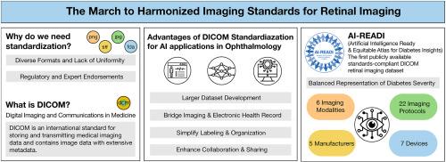
The adoption of standardized imaging protocols in retinal imaging is critical to overcoming challenges posed by fragmented data formats across devices and manufacturers. The lack of standardization hinders clinical interoperability, collaborative research, and the development of artificial intelligence (AI) models that depend on large, high-quality datasets. The Digital Imaging and Communication in Medicine (DICOM) standard offers a robust solution for ensuring interoperability in medical imaging. Although DICOM is widely utilized in radiology and cardiology, its adoption in ophthalmology remains limited. Retinal imaging modalities such as optical coherence tomography (OCT), fundus photography, and OCT angiography (OCTA) have revolutionized retinal disease management but are constrained by proprietary and non-standardized formats.
This review underscores the necessity for harmonized imaging standards in ophthalmology, detailing DICOM standards for retinal imaging including ophthalmic photography (OP), OCT, and OCTA, and their requisite metadata information. Additionally, the potential of DICOM standardization for advancing AI applications in ophthalmology is explored. A notable example is the Artificial Intelligence Ready and Equitable Atlas for Diabetes Insights (AI-READI) dataset, the first publicly available standards-compliant DICOM retinal imaging dataset. This dataset encompasses diverse retinal imaging modalities, including color fundus photography, infrared, autofluorescence, OCT, and OCTA. By leveraging multimodal retinal imaging, AI-READI provides a transformative resource for studying diabetes and its complications, setting a blueprint for future datasets aimed at harmonizing imaging formats and enabling AI-driven breakthroughs in ophthalmology. Our manuscript also addresses challenges in retinal imaging for diabetic patients, retinal imaging-based AI applications for studying diabetes, and potential advancements in retinal imaging standardization.
求助全文
通过发布文献求助,成功后即可免费获取论文全文。
去求助
来源期刊
CiteScore
34.10
自引率
5.10%
发文量
78
期刊介绍:
Progress in Retinal and Eye Research is a Reviews-only journal. By invitation, leading experts write on basic and clinical aspects of the eye in a style appealing to molecular biologists, neuroscientists and physiologists, as well as to vision researchers and ophthalmologists.
The journal covers all aspects of eye research, including topics pertaining to the retina and pigment epithelial layer, cornea, tears, lacrimal glands, aqueous humour, iris, ciliary body, trabeculum, lens, vitreous humour and diseases such as dry-eye, inflammation, keratoconus, corneal dystrophy, glaucoma and cataract.
 求助内容:
求助内容: 应助结果提醒方式:


