Aakriti Tyagi, Disha Mittal, S Bhanoth, Ankita Leekha, Anita K Verma
{"title":"荧光油酸包封ZnSe/CdS/核壳量子点在Balb/c小鼠体内静脉注射后的生物相容性及生物分布评价","authors":"Aakriti Tyagi, Disha Mittal, S Bhanoth, Ankita Leekha, Anita K Verma","doi":"10.34172/bi.30467","DOIUrl":null,"url":null,"abstract":"<p><p></p><p><strong>Introduction: </strong>Quantum dots (QDs) are semiconductor nanocrystals with inherent fluorescence having several advantages over traditional fluorescent probes including their small size (5-10 nm), tunable excitation and emission spectra, ease of surface functionalization, and robust photostability that makes them ideal candidates for <i>in vivo</i> imaging. The behavior of QDs is highly dependent on the surface functionality. <i>In vivo</i> toxicity of QDs in biological systems is the major limitation hindering their translation to clinics.</p><p><strong>Methods: </strong>In the present study, cytotoxicity of water soluble ZnSe/CdS core shell QDs capped with oleic acid was evaluated against human hepatocellular carcinoma cell line-Hep3B, Human Embryonic Kidney cell line-HEK 293 and Ehlrich Ascitic cells-EAC. To assess its <i>in vivo</i> therapeutic efficacy, the initial animal toxicity studies of OA capped ZnSe/ CdS QDs were done in Balb/c mice. Serum stability, pharmacokinetics, biodistribution and γ-scintigraphic imaging were observed in mice after intravenous (<i>i.v</i>) injection of QDs at a dose of 10 nM/kg/200 µL/mice up to 28 days.</p><p><strong>Results: </strong>IC<sub>50</sub> of OA capped QDs against Hep3B was 29.85 µg/mL at 24 hours. QDs toxicity was primarily due to the generation of reactive oxygen species as observed by LDH release in Hep3B cells. Negligible haemolysis indicated that OA capped QDs were biocompatible. OA capped QDs mainly accumulated in the liver and spleen with no retention in kidneys.</p><p><strong>Conclusion: </strong>OA capped ZnSe/ CdS QDs exhibited enhanced anti-cancer efficacy against Hep3B and EAC cell line. Further, minimum accumulation and retention were observed in vital organs in Balb/c mice protecting them from potential adverse side effects, underlining their potential for biomedical applications.</p>","PeriodicalId":48614,"journal":{"name":"Bioimpacts","volume":"15 ","pages":"30467"},"PeriodicalIF":2.2000,"publicationDate":"2025-02-17","publicationTypes":"Journal Article","fieldsOfStudy":null,"isOpenAccess":false,"openAccessPdf":"https://www.ncbi.nlm.nih.gov/pmc/articles/PMC12008253/pdf/","citationCount":"0","resultStr":"{\"title\":\"Assessment of the biocompatibility and biodistribution of fluorescent oleic acid capped ZnSe/CdS/ core shell quantum dots after intravenous injection in Balb/c mice.\",\"authors\":\"Aakriti Tyagi, Disha Mittal, S Bhanoth, Ankita Leekha, Anita K Verma\",\"doi\":\"10.34172/bi.30467\",\"DOIUrl\":null,\"url\":null,\"abstract\":\"<p><p></p><p><strong>Introduction: </strong>Quantum dots (QDs) are semiconductor nanocrystals with inherent fluorescence having several advantages over traditional fluorescent probes including their small size (5-10 nm), tunable excitation and emission spectra, ease of surface functionalization, and robust photostability that makes them ideal candidates for <i>in vivo</i> imaging. The behavior of QDs is highly dependent on the surface functionality. <i>In vivo</i> toxicity of QDs in biological systems is the major limitation hindering their translation to clinics.</p><p><strong>Methods: </strong>In the present study, cytotoxicity of water soluble ZnSe/CdS core shell QDs capped with oleic acid was evaluated against human hepatocellular carcinoma cell line-Hep3B, Human Embryonic Kidney cell line-HEK 293 and Ehlrich Ascitic cells-EAC. To assess its <i>in vivo</i> therapeutic efficacy, the initial animal toxicity studies of OA capped ZnSe/ CdS QDs were done in Balb/c mice. Serum stability, pharmacokinetics, biodistribution and γ-scintigraphic imaging were observed in mice after intravenous (<i>i.v</i>) injection of QDs at a dose of 10 nM/kg/200 µL/mice up to 28 days.</p><p><strong>Results: </strong>IC<sub>50</sub> of OA capped QDs against Hep3B was 29.85 µg/mL at 24 hours. QDs toxicity was primarily due to the generation of reactive oxygen species as observed by LDH release in Hep3B cells. Negligible haemolysis indicated that OA capped QDs were biocompatible. OA capped QDs mainly accumulated in the liver and spleen with no retention in kidneys.</p><p><strong>Conclusion: </strong>OA capped ZnSe/ CdS QDs exhibited enhanced anti-cancer efficacy against Hep3B and EAC cell line. Further, minimum accumulation and retention were observed in vital organs in Balb/c mice protecting them from potential adverse side effects, underlining their potential for biomedical applications.</p>\",\"PeriodicalId\":48614,\"journal\":{\"name\":\"Bioimpacts\",\"volume\":\"15 \",\"pages\":\"30467\"},\"PeriodicalIF\":2.2000,\"publicationDate\":\"2025-02-17\",\"publicationTypes\":\"Journal Article\",\"fieldsOfStudy\":null,\"isOpenAccess\":false,\"openAccessPdf\":\"https://www.ncbi.nlm.nih.gov/pmc/articles/PMC12008253/pdf/\",\"citationCount\":\"0\",\"resultStr\":null,\"platform\":\"Semanticscholar\",\"paperid\":null,\"PeriodicalName\":\"Bioimpacts\",\"FirstCategoryId\":\"5\",\"ListUrlMain\":\"https://doi.org/10.34172/bi.30467\",\"RegionNum\":4,\"RegionCategory\":\"工程技术\",\"ArticlePicture\":[],\"TitleCN\":null,\"AbstractTextCN\":null,\"PMCID\":null,\"EPubDate\":\"2025/1/1 0:00:00\",\"PubModel\":\"eCollection\",\"JCR\":\"Q3\",\"JCRName\":\"PHARMACOLOGY & PHARMACY\",\"Score\":null,\"Total\":0}","platform":"Semanticscholar","paperid":null,"PeriodicalName":"Bioimpacts","FirstCategoryId":"5","ListUrlMain":"https://doi.org/10.34172/bi.30467","RegionNum":4,"RegionCategory":"工程技术","ArticlePicture":[],"TitleCN":null,"AbstractTextCN":null,"PMCID":null,"EPubDate":"2025/1/1 0:00:00","PubModel":"eCollection","JCR":"Q3","JCRName":"PHARMACOLOGY & PHARMACY","Score":null,"Total":0}
Assessment of the biocompatibility and biodistribution of fluorescent oleic acid capped ZnSe/CdS/ core shell quantum dots after intravenous injection in Balb/c mice.
Introduction: Quantum dots (QDs) are semiconductor nanocrystals with inherent fluorescence having several advantages over traditional fluorescent probes including their small size (5-10 nm), tunable excitation and emission spectra, ease of surface functionalization, and robust photostability that makes them ideal candidates for in vivo imaging. The behavior of QDs is highly dependent on the surface functionality. In vivo toxicity of QDs in biological systems is the major limitation hindering their translation to clinics.
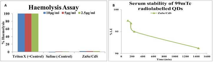
Methods: In the present study, cytotoxicity of water soluble ZnSe/CdS core shell QDs capped with oleic acid was evaluated against human hepatocellular carcinoma cell line-Hep3B, Human Embryonic Kidney cell line-HEK 293 and Ehlrich Ascitic cells-EAC. To assess its in vivo therapeutic efficacy, the initial animal toxicity studies of OA capped ZnSe/ CdS QDs were done in Balb/c mice. Serum stability, pharmacokinetics, biodistribution and γ-scintigraphic imaging were observed in mice after intravenous (i.v) injection of QDs at a dose of 10 nM/kg/200 µL/mice up to 28 days.
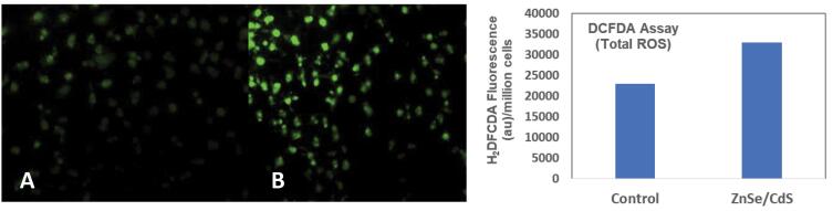
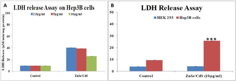
Results: IC50 of OA capped QDs against Hep3B was 29.85 µg/mL at 24 hours. QDs toxicity was primarily due to the generation of reactive oxygen species as observed by LDH release in Hep3B cells. Negligible haemolysis indicated that OA capped QDs were biocompatible. OA capped QDs mainly accumulated in the liver and spleen with no retention in kidneys.
Conclusion: OA capped ZnSe/ CdS QDs exhibited enhanced anti-cancer efficacy against Hep3B and EAC cell line. Further, minimum accumulation and retention were observed in vital organs in Balb/c mice protecting them from potential adverse side effects, underlining their potential for biomedical applications.
BioimpactsPharmacology, Toxicology and Pharmaceutics-Pharmaceutical Science
CiteScore
4.80
自引率
7.70%
发文量
36
审稿时长
5 weeks
期刊介绍:
BioImpacts (BI) is a peer-reviewed multidisciplinary international journal, covering original research articles, reviews, commentaries, hypotheses, methodologies, and visions/reflections dealing with all aspects of biological and biomedical researches at molecular, cellular, functional and translational dimensions.
 求助内容:
求助内容: 应助结果提醒方式:


