{"title":"儿童急性播散性脑脊髓炎血清阴性的性别和年龄差异:来自三级中心的结果和见解。","authors":"Reza Nejad Shahrokh Abadi, Farrokh Seilanian Toosi, Javad Akhoondian, Mehran Beiraghi Toosi, Farah Ashrafzadeh, Mohammadali Nahayati, Shima Shekari, Samaneh Kamali, Shima Imannezhad, Ahmad Sohrab Niazi, Narges Hashemi","doi":"10.22037/ijcn.v19i2.46613","DOIUrl":null,"url":null,"abstract":"<p><strong>Objectives: </strong>Acute disseminated encephalomyelitis (ADEM) is a rapid-onset inflammatory central nervous system (CNS) disorder in children, causing demyelination, encephalopathy, and neurological deficits, often following infections.</p><p><strong>Materials & methods: </strong>This 10-year retrospective study evaluated pediatric patients with seronegative acute disseminated encephalomyelitis (ADEM), focusing on clinical, laboratory, and imaging profiles. The various profiles were assessed to determine age- and/or sex-based differences.</p><p><strong>Results: </strong>The study reviewed 36 patients, with an average age of 6.08 years and predominantly male (61.1%). Clinical presentations included fever, nausea, vomiting, and seizures, with left facial hemiparesis being more common in girls (P-value = 0.023), while abnormal deep tendon reflexes (DTRs) and right-sided pathologies were more common in older patients (P-value < 0.05). Recent laboratory results have revealed differences between peripheral lymphocytes and polymorphonuclear (PMN) cells. Imaging revealed predominantly bilateral lesions, with older patients more likely to show lesions in the right parietal and occipital lobes (P-value = 0.01 and 0.04). Bilateral parietal lobe lesions were significantly correlated with several laboratory findings across the different subgroups. Multivariate logistic regression revealed that these findings were statistically significant in regards to peripheral PMN and lymphocytes in the age category and cerebrospinal fluid (CSF) protein in the gender category (P-value < 0.05). Additionally, girls, particularly those who were older, had significantly higher involvement of the cervical spine (P-value = 0.04 and 0.02).</p><p><strong>Conclusion: </strong>This study reveals age and sex-related differences in the clinical presentation and imaging findings of seronegative pediatric ADEM, showcasing the various demographic factors in patient profiles.</p>","PeriodicalId":14537,"journal":{"name":"Iranian Journal of Child Neurology","volume":"19 2","pages":"77-91"},"PeriodicalIF":0.9000,"publicationDate":"2025-01-01","publicationTypes":"Journal Article","fieldsOfStudy":null,"isOpenAccess":false,"openAccessPdf":"https://www.ncbi.nlm.nih.gov/pmc/articles/PMC11994128/pdf/","citationCount":"0","resultStr":"{\"title\":\"Gender and Age Differences in Seronegative Pediatric Acute Disseminated Encephalomyelitis Profiles: Results and Insights from a Tertiary Center.\",\"authors\":\"Reza Nejad Shahrokh Abadi, Farrokh Seilanian Toosi, Javad Akhoondian, Mehran Beiraghi Toosi, Farah Ashrafzadeh, Mohammadali Nahayati, Shima Shekari, Samaneh Kamali, Shima Imannezhad, Ahmad Sohrab Niazi, Narges Hashemi\",\"doi\":\"10.22037/ijcn.v19i2.46613\",\"DOIUrl\":null,\"url\":null,\"abstract\":\"<p><strong>Objectives: </strong>Acute disseminated encephalomyelitis (ADEM) is a rapid-onset inflammatory central nervous system (CNS) disorder in children, causing demyelination, encephalopathy, and neurological deficits, often following infections.</p><p><strong>Materials & methods: </strong>This 10-year retrospective study evaluated pediatric patients with seronegative acute disseminated encephalomyelitis (ADEM), focusing on clinical, laboratory, and imaging profiles. The various profiles were assessed to determine age- and/or sex-based differences.</p><p><strong>Results: </strong>The study reviewed 36 patients, with an average age of 6.08 years and predominantly male (61.1%). Clinical presentations included fever, nausea, vomiting, and seizures, with left facial hemiparesis being more common in girls (P-value = 0.023), while abnormal deep tendon reflexes (DTRs) and right-sided pathologies were more common in older patients (P-value < 0.05). Recent laboratory results have revealed differences between peripheral lymphocytes and polymorphonuclear (PMN) cells. Imaging revealed predominantly bilateral lesions, with older patients more likely to show lesions in the right parietal and occipital lobes (P-value = 0.01 and 0.04). Bilateral parietal lobe lesions were significantly correlated with several laboratory findings across the different subgroups. Multivariate logistic regression revealed that these findings were statistically significant in regards to peripheral PMN and lymphocytes in the age category and cerebrospinal fluid (CSF) protein in the gender category (P-value < 0.05). Additionally, girls, particularly those who were older, had significantly higher involvement of the cervical spine (P-value = 0.04 and 0.02).</p><p><strong>Conclusion: </strong>This study reveals age and sex-related differences in the clinical presentation and imaging findings of seronegative pediatric ADEM, showcasing the various demographic factors in patient profiles.</p>\",\"PeriodicalId\":14537,\"journal\":{\"name\":\"Iranian Journal of Child Neurology\",\"volume\":\"19 2\",\"pages\":\"77-91\"},\"PeriodicalIF\":0.9000,\"publicationDate\":\"2025-01-01\",\"publicationTypes\":\"Journal Article\",\"fieldsOfStudy\":null,\"isOpenAccess\":false,\"openAccessPdf\":\"https://www.ncbi.nlm.nih.gov/pmc/articles/PMC11994128/pdf/\",\"citationCount\":\"0\",\"resultStr\":null,\"platform\":\"Semanticscholar\",\"paperid\":null,\"PeriodicalName\":\"Iranian Journal of Child Neurology\",\"FirstCategoryId\":\"1085\",\"ListUrlMain\":\"https://doi.org/10.22037/ijcn.v19i2.46613\",\"RegionNum\":0,\"RegionCategory\":null,\"ArticlePicture\":[],\"TitleCN\":null,\"AbstractTextCN\":null,\"PMCID\":null,\"EPubDate\":\"2025/3/11 0:00:00\",\"PubModel\":\"Epub\",\"JCR\":\"Q4\",\"JCRName\":\"CLINICAL NEUROLOGY\",\"Score\":null,\"Total\":0}","platform":"Semanticscholar","paperid":null,"PeriodicalName":"Iranian Journal of Child Neurology","FirstCategoryId":"1085","ListUrlMain":"https://doi.org/10.22037/ijcn.v19i2.46613","RegionNum":0,"RegionCategory":null,"ArticlePicture":[],"TitleCN":null,"AbstractTextCN":null,"PMCID":null,"EPubDate":"2025/3/11 0:00:00","PubModel":"Epub","JCR":"Q4","JCRName":"CLINICAL NEUROLOGY","Score":null,"Total":0}
Gender and Age Differences in Seronegative Pediatric Acute Disseminated Encephalomyelitis Profiles: Results and Insights from a Tertiary Center.
Objectives: Acute disseminated encephalomyelitis (ADEM) is a rapid-onset inflammatory central nervous system (CNS) disorder in children, causing demyelination, encephalopathy, and neurological deficits, often following infections.
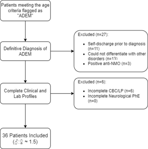
Materials & methods: This 10-year retrospective study evaluated pediatric patients with seronegative acute disseminated encephalomyelitis (ADEM), focusing on clinical, laboratory, and imaging profiles. The various profiles were assessed to determine age- and/or sex-based differences.
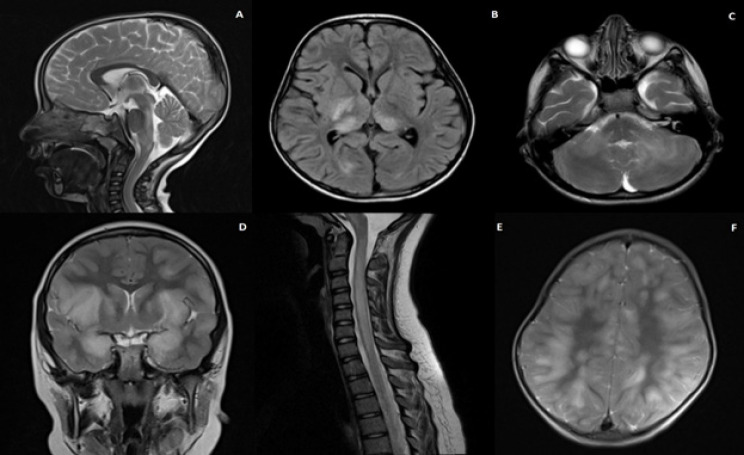
Results: The study reviewed 36 patients, with an average age of 6.08 years and predominantly male (61.1%). Clinical presentations included fever, nausea, vomiting, and seizures, with left facial hemiparesis being more common in girls (P-value = 0.023), while abnormal deep tendon reflexes (DTRs) and right-sided pathologies were more common in older patients (P-value < 0.05). Recent laboratory results have revealed differences between peripheral lymphocytes and polymorphonuclear (PMN) cells. Imaging revealed predominantly bilateral lesions, with older patients more likely to show lesions in the right parietal and occipital lobes (P-value = 0.01 and 0.04). Bilateral parietal lobe lesions were significantly correlated with several laboratory findings across the different subgroups. Multivariate logistic regression revealed that these findings were statistically significant in regards to peripheral PMN and lymphocytes in the age category and cerebrospinal fluid (CSF) protein in the gender category (P-value < 0.05). Additionally, girls, particularly those who were older, had significantly higher involvement of the cervical spine (P-value = 0.04 and 0.02).
Conclusion: This study reveals age and sex-related differences in the clinical presentation and imaging findings of seronegative pediatric ADEM, showcasing the various demographic factors in patient profiles.

 求助内容:
求助内容: 应助结果提醒方式:


