{"title":"显微与全内窥镜下微血管减压治疗面肌痉挛手术效果比较。","authors":"Kyosuke Matsunaga, Norio Ichimasu, Nobuyuki Nakajima, Michihiro Kohno","doi":"10.2176/jns-nmc.2024-0245","DOIUrl":null,"url":null,"abstract":"<p><p>Fully endoscopic microvascular decompression is increasingly being used to treat hemifacial spasm; however, its efficacy must be validated by comparing it with conventional microscopic microvascular decompression. In this study, we aimed to compare the surgical outcomes of microsurgical and endoscopic microvascular decompression for hemifacial spasm and discuss the usefulness and risks of endoscopic treatment. A total of 40 patients with hemifacial spasm were retrospectively evaluated at a single institution between 2016 and 2022, including 33 patients who underwent microscopic microvascular decompression (microvascular decompression group) and 7 patients who underwent fully endoscopic microvascular decompression group, which was chosen for patients with sufficient space in the cerebellopontine cistern for endoscopic manipulation. Statistical analyses of the microvascular decompression group and the endoscopic microvascular decompression group were performed to compare patient background and surgical outcomes. No significant differences in age, sex, or affected side were observed between the 2 groups. At the 6-month follow-up, substantial improvement was observed in more than 85% of the patients in each group. Delayed facial palsy and mild lower cranial nerve palsy, such as hoarseness, were more common in the endoscopic microvascular decompression group than in the microvascular decompression group, although there were no significant differences in the rate of complications between the 2 groups. All complications were alleviated within 3 months after surgery. During endoscopic microvascular decompression, interference between the endoscope and instruments can cause neural damage owing to the limited space along the petrosal surface of the cerebellum. Our results suggest that endoscopic procedures cannot always be used as a substitute for conventional microscopic microvascular decompression.</p>","PeriodicalId":19225,"journal":{"name":"Neurologia medico-chirurgica","volume":" ","pages":"230-238"},"PeriodicalIF":2.3000,"publicationDate":"2025-05-15","publicationTypes":"Journal Article","fieldsOfStudy":null,"isOpenAccess":false,"openAccessPdf":"https://www.ncbi.nlm.nih.gov/pmc/articles/PMC12137054/pdf/","citationCount":"0","resultStr":"{\"title\":\"Comparison of Surgical Outcomes in Microscopic and Fully Endoscopic Microvascular Decompression for Hemifacial Spasm.\",\"authors\":\"Kyosuke Matsunaga, Norio Ichimasu, Nobuyuki Nakajima, Michihiro Kohno\",\"doi\":\"10.2176/jns-nmc.2024-0245\",\"DOIUrl\":null,\"url\":null,\"abstract\":\"<p><p>Fully endoscopic microvascular decompression is increasingly being used to treat hemifacial spasm; however, its efficacy must be validated by comparing it with conventional microscopic microvascular decompression. In this study, we aimed to compare the surgical outcomes of microsurgical and endoscopic microvascular decompression for hemifacial spasm and discuss the usefulness and risks of endoscopic treatment. A total of 40 patients with hemifacial spasm were retrospectively evaluated at a single institution between 2016 and 2022, including 33 patients who underwent microscopic microvascular decompression (microvascular decompression group) and 7 patients who underwent fully endoscopic microvascular decompression group, which was chosen for patients with sufficient space in the cerebellopontine cistern for endoscopic manipulation. Statistical analyses of the microvascular decompression group and the endoscopic microvascular decompression group were performed to compare patient background and surgical outcomes. No significant differences in age, sex, or affected side were observed between the 2 groups. At the 6-month follow-up, substantial improvement was observed in more than 85% of the patients in each group. Delayed facial palsy and mild lower cranial nerve palsy, such as hoarseness, were more common in the endoscopic microvascular decompression group than in the microvascular decompression group, although there were no significant differences in the rate of complications between the 2 groups. All complications were alleviated within 3 months after surgery. During endoscopic microvascular decompression, interference between the endoscope and instruments can cause neural damage owing to the limited space along the petrosal surface of the cerebellum. Our results suggest that endoscopic procedures cannot always be used as a substitute for conventional microscopic microvascular decompression.</p>\",\"PeriodicalId\":19225,\"journal\":{\"name\":\"Neurologia medico-chirurgica\",\"volume\":\" \",\"pages\":\"230-238\"},\"PeriodicalIF\":2.3000,\"publicationDate\":\"2025-05-15\",\"publicationTypes\":\"Journal Article\",\"fieldsOfStudy\":null,\"isOpenAccess\":false,\"openAccessPdf\":\"https://www.ncbi.nlm.nih.gov/pmc/articles/PMC12137054/pdf/\",\"citationCount\":\"0\",\"resultStr\":null,\"platform\":\"Semanticscholar\",\"paperid\":null,\"PeriodicalName\":\"Neurologia medico-chirurgica\",\"FirstCategoryId\":\"3\",\"ListUrlMain\":\"https://doi.org/10.2176/jns-nmc.2024-0245\",\"RegionNum\":4,\"RegionCategory\":\"医学\",\"ArticlePicture\":[],\"TitleCN\":null,\"AbstractTextCN\":null,\"PMCID\":null,\"EPubDate\":\"2025/3/21 0:00:00\",\"PubModel\":\"Epub\",\"JCR\":\"Q2\",\"JCRName\":\"CLINICAL NEUROLOGY\",\"Score\":null,\"Total\":0}","platform":"Semanticscholar","paperid":null,"PeriodicalName":"Neurologia medico-chirurgica","FirstCategoryId":"3","ListUrlMain":"https://doi.org/10.2176/jns-nmc.2024-0245","RegionNum":4,"RegionCategory":"医学","ArticlePicture":[],"TitleCN":null,"AbstractTextCN":null,"PMCID":null,"EPubDate":"2025/3/21 0:00:00","PubModel":"Epub","JCR":"Q2","JCRName":"CLINICAL NEUROLOGY","Score":null,"Total":0}
Comparison of Surgical Outcomes in Microscopic and Fully Endoscopic Microvascular Decompression for Hemifacial Spasm.
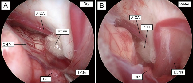
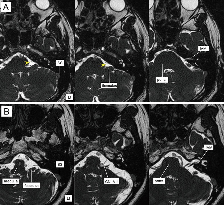
Fully endoscopic microvascular decompression is increasingly being used to treat hemifacial spasm; however, its efficacy must be validated by comparing it with conventional microscopic microvascular decompression. In this study, we aimed to compare the surgical outcomes of microsurgical and endoscopic microvascular decompression for hemifacial spasm and discuss the usefulness and risks of endoscopic treatment. A total of 40 patients with hemifacial spasm were retrospectively evaluated at a single institution between 2016 and 2022, including 33 patients who underwent microscopic microvascular decompression (microvascular decompression group) and 7 patients who underwent fully endoscopic microvascular decompression group, which was chosen for patients with sufficient space in the cerebellopontine cistern for endoscopic manipulation. Statistical analyses of the microvascular decompression group and the endoscopic microvascular decompression group were performed to compare patient background and surgical outcomes. No significant differences in age, sex, or affected side were observed between the 2 groups. At the 6-month follow-up, substantial improvement was observed in more than 85% of the patients in each group. Delayed facial palsy and mild lower cranial nerve palsy, such as hoarseness, were more common in the endoscopic microvascular decompression group than in the microvascular decompression group, although there were no significant differences in the rate of complications between the 2 groups. All complications were alleviated within 3 months after surgery. During endoscopic microvascular decompression, interference between the endoscope and instruments can cause neural damage owing to the limited space along the petrosal surface of the cerebellum. Our results suggest that endoscopic procedures cannot always be used as a substitute for conventional microscopic microvascular decompression.
 求助内容:
求助内容: 应助结果提醒方式:


