Se-Jun Park, Jin-Sung Park, Dong-Ho Kang, Minwook Kang, Kyunghun Jung, Chong-Suh Lee
{"title":"成人脊柱畸形患者尽管实现了理想的矢状面矫正,但近端关节功能衰竭仍在发展:196例低胸骨盆融合手术的危险因素分析","authors":"Se-Jun Park, Jin-Sung Park, Dong-Ho Kang, Minwook Kang, Kyunghun Jung, Chong-Suh Lee","doi":"10.14245/ns.2448734.367","DOIUrl":null,"url":null,"abstract":"<p><strong>Objective: </strong>To identify the risk factors for proximal junctional failure (PJF) after adult spinal deformity (ASD) surgery despite ideal sagittal correction according to age-adjusted alignment target.</p><p><strong>Methods: </strong>The study included patients who underwent low thoracic to pelvic fusion for ASD and obtained ideal correction according to age-adjusted pelvic incidence minus lumbar lordosis. PJF was defined either radiographically as a proximal junctional angle (PJA) of >28° plus a difference in PJA of >22° or clinically as revision surgery for proximal junctional complications. Clinical and radiographic variables were assessed to identify the risk factors for PJF.</p><p><strong>Results: </strong>The final study cohort consisted of 196 patients, of whom 170 were women (86.7%), with an average age of 68.3 years. During mean follow-up duration of 45.9 months, PJF occurred in 43 patients (21.9%). Multivariate logistic regression analysis revealed that old age (odds ratio [OR], 1.063; 95% confidence interval [CI], 1.001-1.129; p=0.046), large preoperative sagittal vertical axis (OR, 1.007; 95% CI, 1.001-1.013; p=0.024), nonuse of a transverse process (TP) hook (OR, 5.556; 95% CI, 1.205-19.621; p=0.028), and high lumbar distribution index (LDI) (OR, 1.136; 95% CI, 1.109-1.164; p<0.001) were significant risk factors for PJF development.</p><p><strong>Conclusion: </strong>A sizeable proportion of patients (21.9%) developed PJF despite achieving ideal sagittal correction. Using TP hooks with avoiding excessive LDI can be helpful to further mitigate the risk of PJF development in this patient group.</p>","PeriodicalId":19269,"journal":{"name":"Neurospine","volume":"21 4","pages":"1080-1090"},"PeriodicalIF":3.6000,"publicationDate":"2024-12-01","publicationTypes":"Journal Article","fieldsOfStudy":null,"isOpenAccess":false,"openAccessPdf":"https://www.ncbi.nlm.nih.gov/pmc/articles/PMC11744533/pdf/","citationCount":"0","resultStr":"{\"title\":\"Proximal Junctional Failure Development Despite Achieving Ideal Sagittal Correction According to Age-Adjusted Alignment Target in Patients With Adult Spinal Deformity: Risk Factor Analysis of 196 Cases Undergoing Low Thoracic to Pelvic Fusion.\",\"authors\":\"Se-Jun Park, Jin-Sung Park, Dong-Ho Kang, Minwook Kang, Kyunghun Jung, Chong-Suh Lee\",\"doi\":\"10.14245/ns.2448734.367\",\"DOIUrl\":null,\"url\":null,\"abstract\":\"<p><strong>Objective: </strong>To identify the risk factors for proximal junctional failure (PJF) after adult spinal deformity (ASD) surgery despite ideal sagittal correction according to age-adjusted alignment target.</p><p><strong>Methods: </strong>The study included patients who underwent low thoracic to pelvic fusion for ASD and obtained ideal correction according to age-adjusted pelvic incidence minus lumbar lordosis. PJF was defined either radiographically as a proximal junctional angle (PJA) of >28° plus a difference in PJA of >22° or clinically as revision surgery for proximal junctional complications. Clinical and radiographic variables were assessed to identify the risk factors for PJF.</p><p><strong>Results: </strong>The final study cohort consisted of 196 patients, of whom 170 were women (86.7%), with an average age of 68.3 years. During mean follow-up duration of 45.9 months, PJF occurred in 43 patients (21.9%). Multivariate logistic regression analysis revealed that old age (odds ratio [OR], 1.063; 95% confidence interval [CI], 1.001-1.129; p=0.046), large preoperative sagittal vertical axis (OR, 1.007; 95% CI, 1.001-1.013; p=0.024), nonuse of a transverse process (TP) hook (OR, 5.556; 95% CI, 1.205-19.621; p=0.028), and high lumbar distribution index (LDI) (OR, 1.136; 95% CI, 1.109-1.164; p<0.001) were significant risk factors for PJF development.</p><p><strong>Conclusion: </strong>A sizeable proportion of patients (21.9%) developed PJF despite achieving ideal sagittal correction. Using TP hooks with avoiding excessive LDI can be helpful to further mitigate the risk of PJF development in this patient group.</p>\",\"PeriodicalId\":19269,\"journal\":{\"name\":\"Neurospine\",\"volume\":\"21 4\",\"pages\":\"1080-1090\"},\"PeriodicalIF\":3.6000,\"publicationDate\":\"2024-12-01\",\"publicationTypes\":\"Journal Article\",\"fieldsOfStudy\":null,\"isOpenAccess\":false,\"openAccessPdf\":\"https://www.ncbi.nlm.nih.gov/pmc/articles/PMC11744533/pdf/\",\"citationCount\":\"0\",\"resultStr\":null,\"platform\":\"Semanticscholar\",\"paperid\":null,\"PeriodicalName\":\"Neurospine\",\"FirstCategoryId\":\"3\",\"ListUrlMain\":\"https://doi.org/10.14245/ns.2448734.367\",\"RegionNum\":2,\"RegionCategory\":\"医学\",\"ArticlePicture\":[],\"TitleCN\":null,\"AbstractTextCN\":null,\"PMCID\":null,\"EPubDate\":\"2024/12/31 0:00:00\",\"PubModel\":\"Epub\",\"JCR\":\"Q1\",\"JCRName\":\"CLINICAL NEUROLOGY\",\"Score\":null,\"Total\":0}","platform":"Semanticscholar","paperid":null,"PeriodicalName":"Neurospine","FirstCategoryId":"3","ListUrlMain":"https://doi.org/10.14245/ns.2448734.367","RegionNum":2,"RegionCategory":"医学","ArticlePicture":[],"TitleCN":null,"AbstractTextCN":null,"PMCID":null,"EPubDate":"2024/12/31 0:00:00","PubModel":"Epub","JCR":"Q1","JCRName":"CLINICAL NEUROLOGY","Score":null,"Total":0}
Proximal Junctional Failure Development Despite Achieving Ideal Sagittal Correction According to Age-Adjusted Alignment Target in Patients With Adult Spinal Deformity: Risk Factor Analysis of 196 Cases Undergoing Low Thoracic to Pelvic Fusion.
Objective: To identify the risk factors for proximal junctional failure (PJF) after adult spinal deformity (ASD) surgery despite ideal sagittal correction according to age-adjusted alignment target.
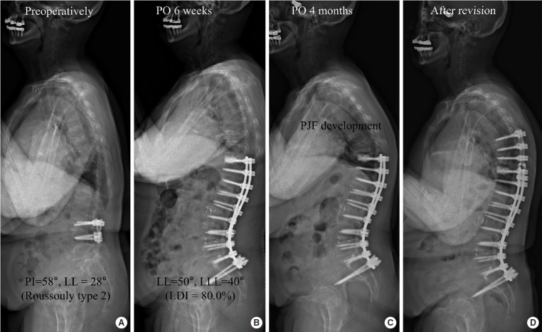
Methods: The study included patients who underwent low thoracic to pelvic fusion for ASD and obtained ideal correction according to age-adjusted pelvic incidence minus lumbar lordosis. PJF was defined either radiographically as a proximal junctional angle (PJA) of >28° plus a difference in PJA of >22° or clinically as revision surgery for proximal junctional complications. Clinical and radiographic variables were assessed to identify the risk factors for PJF.
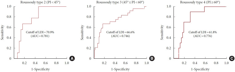
Results: The final study cohort consisted of 196 patients, of whom 170 were women (86.7%), with an average age of 68.3 years. During mean follow-up duration of 45.9 months, PJF occurred in 43 patients (21.9%). Multivariate logistic regression analysis revealed that old age (odds ratio [OR], 1.063; 95% confidence interval [CI], 1.001-1.129; p=0.046), large preoperative sagittal vertical axis (OR, 1.007; 95% CI, 1.001-1.013; p=0.024), nonuse of a transverse process (TP) hook (OR, 5.556; 95% CI, 1.205-19.621; p=0.028), and high lumbar distribution index (LDI) (OR, 1.136; 95% CI, 1.109-1.164; p<0.001) were significant risk factors for PJF development.
Conclusion: A sizeable proportion of patients (21.9%) developed PJF despite achieving ideal sagittal correction. Using TP hooks with avoiding excessive LDI can be helpful to further mitigate the risk of PJF development in this patient group.
 求助内容:
求助内容: 应助结果提醒方式:


