Rofat Askoro, Kota Kagawa, Go Seyama, Akitake Okamura, Akira Hashizume, Tae Onari, Yutaka Hirokawa, Koji Iida, Nobutaka Horie
{"title":"磁共振成像阴性局灶性难治性癫痫的四种无创术前方式联合预测癫痫发作结局。","authors":"Rofat Askoro, Kota Kagawa, Go Seyama, Akitake Okamura, Akira Hashizume, Tae Onari, Yutaka Hirokawa, Koji Iida, Nobutaka Horie","doi":"10.2176/jns-nmc.2024-0194","DOIUrl":null,"url":null,"abstract":"<p><p>In focal epilepsy cases, precise identification and resection of the epileptogenic zone increase the likelihood of achieving a seizure-free outcome. Nevertheless, localizing the source of epilepsy in magnetic resonance imaging-negative epilepsy cases presents significant challenges for clinicians. In this study, we evaluated the diagnostic efficacy and impact on the seizure outcome by using 4 noninvasive modalities, including scalp video electroencephalography, magnetoencephalography, fluorodeoxyglucose-positron emission tomography, and iomazenil single-photon emission computed tomography, in a cohort of patients with magnetic resonance imaging-negative focal epilepsy who underwent resective surgery. The concordance status of each modality was assessed relative to the lobar resection area, and surgical outcome was assessed by Engel Classification at least 1 year after surgery. Comparison and diagnostic analyses were calculated for each individual and all possible combinations of scalp video electroencephalography, magnetoencephalography, fluorodeoxyglucose-positron emission tomography, and single-photon emission computed tomography with respect to Engel class I outcome. Eighteen patients (66.6%, 18/27) had Engel class I outcomes. Patients with at least 2 concordant modalities were associated with Engel class I outcome (p = 0.0262). For individual modality, fluorodeoxyglucose-positron emission tomography achieved the highest yield of sensitivity (72.2%) compared to scalp video electroencephalography, magnetoencephalography, and single-photon emission computed tomography (50.0%, 61.1%, and 61.6%, respectively). Scalp video electroencephalography, magnetoencephalography, and single-photon emission computed tomography showed similar specificities of 77.7%, while fluorodeoxyglucose-positron emission tomography showed a specificity of 55.5%. Combined modalities were able to achieve the highest sensitivity of 83.3% when there were at least 2 concordant modalities and a specificity of 100% with various multiple combinations. Our study showed that lobar concordance from multiple modalities increases the sensitivity and specificity for a seizure-free outcome in magnetic resonance imaging-negative focal epilepsy patients who underwent resective surgery.</p>","PeriodicalId":19225,"journal":{"name":"Neurologia medico-chirurgica","volume":" ","pages":"81-91"},"PeriodicalIF":2.3000,"publicationDate":"2025-02-15","publicationTypes":"Journal Article","fieldsOfStudy":null,"isOpenAccess":false,"openAccessPdf":"https://www.ncbi.nlm.nih.gov/pmc/articles/PMC11891146/pdf/","citationCount":"0","resultStr":"{\"title\":\"Prediction of Seizure Outcome Using Combinations of Four Noninvasive Presurgical Modalities in Magnetic Resonance Imaging-negative Focal Intractable Epilepsy.\",\"authors\":\"Rofat Askoro, Kota Kagawa, Go Seyama, Akitake Okamura, Akira Hashizume, Tae Onari, Yutaka Hirokawa, Koji Iida, Nobutaka Horie\",\"doi\":\"10.2176/jns-nmc.2024-0194\",\"DOIUrl\":null,\"url\":null,\"abstract\":\"<p><p>In focal epilepsy cases, precise identification and resection of the epileptogenic zone increase the likelihood of achieving a seizure-free outcome. Nevertheless, localizing the source of epilepsy in magnetic resonance imaging-negative epilepsy cases presents significant challenges for clinicians. In this study, we evaluated the diagnostic efficacy and impact on the seizure outcome by using 4 noninvasive modalities, including scalp video electroencephalography, magnetoencephalography, fluorodeoxyglucose-positron emission tomography, and iomazenil single-photon emission computed tomography, in a cohort of patients with magnetic resonance imaging-negative focal epilepsy who underwent resective surgery. The concordance status of each modality was assessed relative to the lobar resection area, and surgical outcome was assessed by Engel Classification at least 1 year after surgery. Comparison and diagnostic analyses were calculated for each individual and all possible combinations of scalp video electroencephalography, magnetoencephalography, fluorodeoxyglucose-positron emission tomography, and single-photon emission computed tomography with respect to Engel class I outcome. Eighteen patients (66.6%, 18/27) had Engel class I outcomes. Patients with at least 2 concordant modalities were associated with Engel class I outcome (p = 0.0262). For individual modality, fluorodeoxyglucose-positron emission tomography achieved the highest yield of sensitivity (72.2%) compared to scalp video electroencephalography, magnetoencephalography, and single-photon emission computed tomography (50.0%, 61.1%, and 61.6%, respectively). Scalp video electroencephalography, magnetoencephalography, and single-photon emission computed tomography showed similar specificities of 77.7%, while fluorodeoxyglucose-positron emission tomography showed a specificity of 55.5%. Combined modalities were able to achieve the highest sensitivity of 83.3% when there were at least 2 concordant modalities and a specificity of 100% with various multiple combinations. Our study showed that lobar concordance from multiple modalities increases the sensitivity and specificity for a seizure-free outcome in magnetic resonance imaging-negative focal epilepsy patients who underwent resective surgery.</p>\",\"PeriodicalId\":19225,\"journal\":{\"name\":\"Neurologia medico-chirurgica\",\"volume\":\" \",\"pages\":\"81-91\"},\"PeriodicalIF\":2.3000,\"publicationDate\":\"2025-02-15\",\"publicationTypes\":\"Journal Article\",\"fieldsOfStudy\":null,\"isOpenAccess\":false,\"openAccessPdf\":\"https://www.ncbi.nlm.nih.gov/pmc/articles/PMC11891146/pdf/\",\"citationCount\":\"0\",\"resultStr\":null,\"platform\":\"Semanticscholar\",\"paperid\":null,\"PeriodicalName\":\"Neurologia medico-chirurgica\",\"FirstCategoryId\":\"3\",\"ListUrlMain\":\"https://doi.org/10.2176/jns-nmc.2024-0194\",\"RegionNum\":4,\"RegionCategory\":\"医学\",\"ArticlePicture\":[],\"TitleCN\":null,\"AbstractTextCN\":null,\"PMCID\":null,\"EPubDate\":\"2024/12/10 0:00:00\",\"PubModel\":\"Epub\",\"JCR\":\"Q2\",\"JCRName\":\"CLINICAL NEUROLOGY\",\"Score\":null,\"Total\":0}","platform":"Semanticscholar","paperid":null,"PeriodicalName":"Neurologia medico-chirurgica","FirstCategoryId":"3","ListUrlMain":"https://doi.org/10.2176/jns-nmc.2024-0194","RegionNum":4,"RegionCategory":"医学","ArticlePicture":[],"TitleCN":null,"AbstractTextCN":null,"PMCID":null,"EPubDate":"2024/12/10 0:00:00","PubModel":"Epub","JCR":"Q2","JCRName":"CLINICAL NEUROLOGY","Score":null,"Total":0}
Prediction of Seizure Outcome Using Combinations of Four Noninvasive Presurgical Modalities in Magnetic Resonance Imaging-negative Focal Intractable Epilepsy.
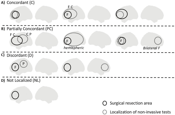
In focal epilepsy cases, precise identification and resection of the epileptogenic zone increase the likelihood of achieving a seizure-free outcome. Nevertheless, localizing the source of epilepsy in magnetic resonance imaging-negative epilepsy cases presents significant challenges for clinicians. In this study, we evaluated the diagnostic efficacy and impact on the seizure outcome by using 4 noninvasive modalities, including scalp video electroencephalography, magnetoencephalography, fluorodeoxyglucose-positron emission tomography, and iomazenil single-photon emission computed tomography, in a cohort of patients with magnetic resonance imaging-negative focal epilepsy who underwent resective surgery. The concordance status of each modality was assessed relative to the lobar resection area, and surgical outcome was assessed by Engel Classification at least 1 year after surgery. Comparison and diagnostic analyses were calculated for each individual and all possible combinations of scalp video electroencephalography, magnetoencephalography, fluorodeoxyglucose-positron emission tomography, and single-photon emission computed tomography with respect to Engel class I outcome. Eighteen patients (66.6%, 18/27) had Engel class I outcomes. Patients with at least 2 concordant modalities were associated with Engel class I outcome (p = 0.0262). For individual modality, fluorodeoxyglucose-positron emission tomography achieved the highest yield of sensitivity (72.2%) compared to scalp video electroencephalography, magnetoencephalography, and single-photon emission computed tomography (50.0%, 61.1%, and 61.6%, respectively). Scalp video electroencephalography, magnetoencephalography, and single-photon emission computed tomography showed similar specificities of 77.7%, while fluorodeoxyglucose-positron emission tomography showed a specificity of 55.5%. Combined modalities were able to achieve the highest sensitivity of 83.3% when there were at least 2 concordant modalities and a specificity of 100% with various multiple combinations. Our study showed that lobar concordance from multiple modalities increases the sensitivity and specificity for a seizure-free outcome in magnetic resonance imaging-negative focal epilepsy patients who underwent resective surgery.
 求助内容:
求助内容: 应助结果提醒方式:


