{"title":"使用短波红外持续发光的硅基和砷化镓基成像设备的深层组织近红外-II 生物成像性能","authors":"Yafei Chen, Simona Spinelli and Zhengwei Pan","doi":"10.1039/D4TC01323A","DOIUrl":null,"url":null,"abstract":"<p >Bioimaging in the second near-infrared (NIR-II; 950–1700 nm) window, employing InGaAs-based cameras, exhibits superior clarity and resolution in visualizing shallow structures (<3 mm), yet it falls short in effectively imaging deep-tissue features. The subpar performance of deep-tissue NIR-II imaging is largely attributed to shortcomings in imaging contrast agents, primarily concerning their luminescence brightness and wavelengths, with minimal consideration given to deficiencies in InGaAs cameras. Here, we use a MgGeO<small><sub>3</sub></small>:Yb<small><sup>3+</sup></small> short-wave infrared (SWIR) persistent luminescent phosphor emitting at 950–1100 nm as a contrast agent to assess the deep-tissue bioimaging capabilities of both an InGaAs camera and a Si CCD camera under identical imaging conditions in thick chicken breast tissues (5–20 mm), thick mice bodies (10–20 mm), and internal mice organs (gastrointestinal tracts, lungs, and livers). Despite the significantly higher quantum efficiency of the InGaAs camera (∼80–85%) compared to the Si camera (∼5–30%) in detecting 950–1100 nm SWIR light, the former exhibits notably inferior performance overall in imaging deep-tissue features, particularly in scenarios with faint imaging signals, attributable to the pronounced interference of its inherently high dark current. Nonetheless, when provided with sufficiently intense SWIR imaging signals, the InGaAs camera outperforms the Si camera in terms of clarity, even in chicken tissues of 10 mm thickness and in the stomachs of mice.</p>","PeriodicalId":84,"journal":{"name":"Journal of Materials Chemistry C","volume":" 21","pages":" 7542-7551"},"PeriodicalIF":5.1000,"publicationDate":"2024-05-02","publicationTypes":"Journal Article","fieldsOfStudy":null,"isOpenAccess":false,"openAccessPdf":"","citationCount":"0","resultStr":"{\"title\":\"Deep-tissue NIR-II bioimaging performance of Si-based and InGaAs-based imaging devices using short-wave infrared persistent luminescence†\",\"authors\":\"Yafei Chen, Simona Spinelli and Zhengwei Pan\",\"doi\":\"10.1039/D4TC01323A\",\"DOIUrl\":null,\"url\":null,\"abstract\":\"<p >Bioimaging in the second near-infrared (NIR-II; 950–1700 nm) window, employing InGaAs-based cameras, exhibits superior clarity and resolution in visualizing shallow structures (<3 mm), yet it falls short in effectively imaging deep-tissue features. The subpar performance of deep-tissue NIR-II imaging is largely attributed to shortcomings in imaging contrast agents, primarily concerning their luminescence brightness and wavelengths, with minimal consideration given to deficiencies in InGaAs cameras. Here, we use a MgGeO<small><sub>3</sub></small>:Yb<small><sup>3+</sup></small> short-wave infrared (SWIR) persistent luminescent phosphor emitting at 950–1100 nm as a contrast agent to assess the deep-tissue bioimaging capabilities of both an InGaAs camera and a Si CCD camera under identical imaging conditions in thick chicken breast tissues (5–20 mm), thick mice bodies (10–20 mm), and internal mice organs (gastrointestinal tracts, lungs, and livers). Despite the significantly higher quantum efficiency of the InGaAs camera (∼80–85%) compared to the Si camera (∼5–30%) in detecting 950–1100 nm SWIR light, the former exhibits notably inferior performance overall in imaging deep-tissue features, particularly in scenarios with faint imaging signals, attributable to the pronounced interference of its inherently high dark current. Nonetheless, when provided with sufficiently intense SWIR imaging signals, the InGaAs camera outperforms the Si camera in terms of clarity, even in chicken tissues of 10 mm thickness and in the stomachs of mice.</p>\",\"PeriodicalId\":84,\"journal\":{\"name\":\"Journal of Materials Chemistry C\",\"volume\":\" 21\",\"pages\":\" 7542-7551\"},\"PeriodicalIF\":5.1000,\"publicationDate\":\"2024-05-02\",\"publicationTypes\":\"Journal Article\",\"fieldsOfStudy\":null,\"isOpenAccess\":false,\"openAccessPdf\":\"\",\"citationCount\":\"0\",\"resultStr\":null,\"platform\":\"Semanticscholar\",\"paperid\":null,\"PeriodicalName\":\"Journal of Materials Chemistry C\",\"FirstCategoryId\":\"1\",\"ListUrlMain\":\"https://pubs.rsc.org/en/content/articlelanding/2024/tc/d4tc01323a\",\"RegionNum\":2,\"RegionCategory\":\"材料科学\",\"ArticlePicture\":[],\"TitleCN\":null,\"AbstractTextCN\":null,\"PMCID\":null,\"EPubDate\":\"\",\"PubModel\":\"\",\"JCR\":\"Q2\",\"JCRName\":\"MATERIALS SCIENCE, MULTIDISCIPLINARY\",\"Score\":null,\"Total\":0}","platform":"Semanticscholar","paperid":null,"PeriodicalName":"Journal of Materials Chemistry C","FirstCategoryId":"1","ListUrlMain":"https://pubs.rsc.org/en/content/articlelanding/2024/tc/d4tc01323a","RegionNum":2,"RegionCategory":"材料科学","ArticlePicture":[],"TitleCN":null,"AbstractTextCN":null,"PMCID":null,"EPubDate":"","PubModel":"","JCR":"Q2","JCRName":"MATERIALS SCIENCE, MULTIDISCIPLINARY","Score":null,"Total":0}
Deep-tissue NIR-II bioimaging performance of Si-based and InGaAs-based imaging devices using short-wave infrared persistent luminescence†
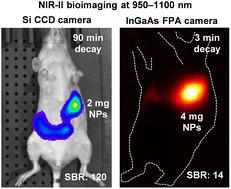
Bioimaging in the second near-infrared (NIR-II; 950–1700 nm) window, employing InGaAs-based cameras, exhibits superior clarity and resolution in visualizing shallow structures (<3 mm), yet it falls short in effectively imaging deep-tissue features. The subpar performance of deep-tissue NIR-II imaging is largely attributed to shortcomings in imaging contrast agents, primarily concerning their luminescence brightness and wavelengths, with minimal consideration given to deficiencies in InGaAs cameras. Here, we use a MgGeO3:Yb3+ short-wave infrared (SWIR) persistent luminescent phosphor emitting at 950–1100 nm as a contrast agent to assess the deep-tissue bioimaging capabilities of both an InGaAs camera and a Si CCD camera under identical imaging conditions in thick chicken breast tissues (5–20 mm), thick mice bodies (10–20 mm), and internal mice organs (gastrointestinal tracts, lungs, and livers). Despite the significantly higher quantum efficiency of the InGaAs camera (∼80–85%) compared to the Si camera (∼5–30%) in detecting 950–1100 nm SWIR light, the former exhibits notably inferior performance overall in imaging deep-tissue features, particularly in scenarios with faint imaging signals, attributable to the pronounced interference of its inherently high dark current. Nonetheless, when provided with sufficiently intense SWIR imaging signals, the InGaAs camera outperforms the Si camera in terms of clarity, even in chicken tissues of 10 mm thickness and in the stomachs of mice.
期刊介绍:
The Journal of Materials Chemistry is divided into three distinct sections, A, B, and C, each catering to specific applications of the materials under study:
Journal of Materials Chemistry A focuses primarily on materials intended for applications in energy and sustainability.
Journal of Materials Chemistry B specializes in materials designed for applications in biology and medicine.
Journal of Materials Chemistry C is dedicated to materials suitable for applications in optical, magnetic, and electronic devices.
Example topic areas within the scope of Journal of Materials Chemistry C are listed below. This list is neither exhaustive nor exclusive.
Bioelectronics
Conductors
Detectors
Dielectrics
Displays
Ferroelectrics
Lasers
LEDs
Lighting
Liquid crystals
Memory
Metamaterials
Multiferroics
Photonics
Photovoltaics
Semiconductors
Sensors
Single molecule conductors
Spintronics
Superconductors
Thermoelectrics
Topological insulators
Transistors

 求助内容:
求助内容: 应助结果提醒方式:


