Haya A. Homsi MD, MPH, Calvin Knapp III MD, Shruti Agrawal MD, Shweta Bhavsar MD, Jennifer S. Ko MD, PhD, Steven D. Billings MD, Shira Ronen MD
{"title":"皮肤晶体贮积组织细胞增生症:系列病例及文献综述","authors":"Haya A. Homsi MD, MPH, Calvin Knapp III MD, Shruti Agrawal MD, Shweta Bhavsar MD, Jennifer S. Ko MD, PhD, Steven D. Billings MD, Shira Ronen MD","doi":"10.1111/cup.14625","DOIUrl":null,"url":null,"abstract":"<p>Crystal-storing histiocytosis (CSH) is a rare condition in which crystals accumulate in the cytoplasm of histiocytes and is usually associated with a lymphoplasmacytic neoplasm. Cutaneous CSH is extraordinarily rare and limited to case reports in the literature. We report two cases of this disease with cutaneous involvement. Case 1 was a 65-year-old male with a 4-month history of a pruritic eruption that started as a solitary pink to skin-colored indurated plaque on the anterior neck before progressing to involve the whole neck, chest wall, and face. Case 2 was a 54-year-old woman with a history of unspecified “lymphoma” who presented with a soft nodule on the forearm. Biopsies from both cases had similar findings and showed a proliferation of epithelioid cells with pink cytoplasm and intracellular crystalline structures infiltrating the dermis and subcutaneous fat. In the first case, the cells were positive for CD43, CD45, CD68, and IgG kappa, and in the second case, the crystals were positive for IgG lambda. Based on these findings, the patients were diagnosed with cutaneous CSH. We highlight this rare diagnosis and the importance of investigating an underlying lymphoplasmacytic neoplasm.</p>","PeriodicalId":15407,"journal":{"name":"Journal of Cutaneous Pathology","volume":null,"pages":null},"PeriodicalIF":1.6000,"publicationDate":"2024-04-12","publicationTypes":"Journal Article","fieldsOfStudy":null,"isOpenAccess":false,"openAccessPdf":"https://onlinelibrary.wiley.com/doi/epdf/10.1111/cup.14625","citationCount":"0","resultStr":"{\"title\":\"Cutaneous crystal storing histiocytosis: A case series with review of literature\",\"authors\":\"Haya A. Homsi MD, MPH, Calvin Knapp III MD, Shruti Agrawal MD, Shweta Bhavsar MD, Jennifer S. Ko MD, PhD, Steven D. Billings MD, Shira Ronen MD\",\"doi\":\"10.1111/cup.14625\",\"DOIUrl\":null,\"url\":null,\"abstract\":\"<p>Crystal-storing histiocytosis (CSH) is a rare condition in which crystals accumulate in the cytoplasm of histiocytes and is usually associated with a lymphoplasmacytic neoplasm. Cutaneous CSH is extraordinarily rare and limited to case reports in the literature. We report two cases of this disease with cutaneous involvement. Case 1 was a 65-year-old male with a 4-month history of a pruritic eruption that started as a solitary pink to skin-colored indurated plaque on the anterior neck before progressing to involve the whole neck, chest wall, and face. Case 2 was a 54-year-old woman with a history of unspecified “lymphoma” who presented with a soft nodule on the forearm. Biopsies from both cases had similar findings and showed a proliferation of epithelioid cells with pink cytoplasm and intracellular crystalline structures infiltrating the dermis and subcutaneous fat. In the first case, the cells were positive for CD43, CD45, CD68, and IgG kappa, and in the second case, the crystals were positive for IgG lambda. Based on these findings, the patients were diagnosed with cutaneous CSH. We highlight this rare diagnosis and the importance of investigating an underlying lymphoplasmacytic neoplasm.</p>\",\"PeriodicalId\":15407,\"journal\":{\"name\":\"Journal of Cutaneous Pathology\",\"volume\":null,\"pages\":null},\"PeriodicalIF\":1.6000,\"publicationDate\":\"2024-04-12\",\"publicationTypes\":\"Journal Article\",\"fieldsOfStudy\":null,\"isOpenAccess\":false,\"openAccessPdf\":\"https://onlinelibrary.wiley.com/doi/epdf/10.1111/cup.14625\",\"citationCount\":\"0\",\"resultStr\":null,\"platform\":\"Semanticscholar\",\"paperid\":null,\"PeriodicalName\":\"Journal of Cutaneous Pathology\",\"FirstCategoryId\":\"3\",\"ListUrlMain\":\"https://onlinelibrary.wiley.com/doi/10.1111/cup.14625\",\"RegionNum\":4,\"RegionCategory\":\"医学\",\"ArticlePicture\":[],\"TitleCN\":null,\"AbstractTextCN\":null,\"PMCID\":null,\"EPubDate\":\"\",\"PubModel\":\"\",\"JCR\":\"Q3\",\"JCRName\":\"DERMATOLOGY\",\"Score\":null,\"Total\":0}","platform":"Semanticscholar","paperid":null,"PeriodicalName":"Journal of Cutaneous Pathology","FirstCategoryId":"3","ListUrlMain":"https://onlinelibrary.wiley.com/doi/10.1111/cup.14625","RegionNum":4,"RegionCategory":"医学","ArticlePicture":[],"TitleCN":null,"AbstractTextCN":null,"PMCID":null,"EPubDate":"","PubModel":"","JCR":"Q3","JCRName":"DERMATOLOGY","Score":null,"Total":0}
Cutaneous crystal storing histiocytosis: A case series with review of literature
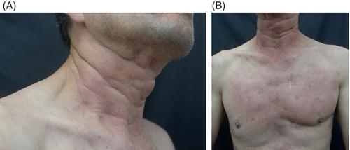
Crystal-storing histiocytosis (CSH) is a rare condition in which crystals accumulate in the cytoplasm of histiocytes and is usually associated with a lymphoplasmacytic neoplasm. Cutaneous CSH is extraordinarily rare and limited to case reports in the literature. We report two cases of this disease with cutaneous involvement. Case 1 was a 65-year-old male with a 4-month history of a pruritic eruption that started as a solitary pink to skin-colored indurated plaque on the anterior neck before progressing to involve the whole neck, chest wall, and face. Case 2 was a 54-year-old woman with a history of unspecified “lymphoma” who presented with a soft nodule on the forearm. Biopsies from both cases had similar findings and showed a proliferation of epithelioid cells with pink cytoplasm and intracellular crystalline structures infiltrating the dermis and subcutaneous fat. In the first case, the cells were positive for CD43, CD45, CD68, and IgG kappa, and in the second case, the crystals were positive for IgG lambda. Based on these findings, the patients were diagnosed with cutaneous CSH. We highlight this rare diagnosis and the importance of investigating an underlying lymphoplasmacytic neoplasm.
期刊介绍:
Journal of Cutaneous Pathology publishes manuscripts broadly relevant to diseases of the skin and mucosae, with the aims of advancing scientific knowledge regarding dermatopathology and enhancing the communication between clinical practitioners and research scientists. Original scientific manuscripts on diagnostic and experimental cutaneous pathology are especially desirable. Timely, pertinent review articles also will be given high priority. Manuscripts based on light, fluorescence, and electron microscopy, histochemistry, immunology, molecular biology, and genetics, as well as allied sciences, are all welcome, provided their principal focus is on cutaneous pathology. Publication time will be kept as short as possible, ensuring that articles will be quickly available to all interested in this speciality.

 求助内容:
求助内容: 应助结果提醒方式:


