Brenna C. Li, Scott Hammond, Anil V. Parwani, Rulong Shen
{"title":"人工智能算法准确评估转移性乳腺癌细胞学标本中的雌激素受体免疫组化:试点研究。","authors":"Brenna C. Li, Scott Hammond, Anil V. Parwani, Rulong Shen","doi":"10.1111/cyt.13373","DOIUrl":null,"url":null,"abstract":"<div>\n \n \n <section>\n \n <h3> Objective</h3>\n \n <p>The Visiopharm artificial intelligence (AI) algorithm for oestrogen receptor (ER) immunohistochemistry (IHC) in whole slide images (WSIs) has been successfully validated in surgical pathology. This study aimed to assess its efficacy in cytology specimens.</p>\n </section>\n \n <section>\n \n <h3> Methods</h3>\n \n <p>The study cohort comprised 105 consecutive cytology specimens with metastatic breast carcinoma. ER IHC WSIs were seamlessly integrated into the Visiopharm platform from the Image Management System (IMS) during our routine digital workflow, and an AI algorithm was employed for analysis. ER AI scores were compared with pathologists' manual consensus scores. Optimization steps were implemented and evaluated to reduce discordance.</p>\n </section>\n \n <section>\n \n <h3> Results</h3>\n \n <p>The overall concordance between pathologists' scores and AI scores was excellent (99/105, 94.3%). Six cases exhibited discordant results, including two false-negative (FN) cases due to abundant histiocytes incorrectly counted as negatively stained tumour cells by AI, two FN cases owing to weak staining, and two false-positive (FP) cases where pigmented macrophages were erroneously counted as positively stained tumour cells by AI. The Pearson correlation coefficient of ER-positive percentages between pathologists' and AI scores was 0.8483. Optimization steps, such as lowering the cut-off threshold and additional training using higher input magnification, significantly improved accuracy.</p>\n </section>\n \n <section>\n \n <h3> Conclusions</h3>\n \n <p>The automated ER AI algorithm demonstrated excellent concordance with pathologists' assessments and accurately differentiated ER-positive from ER-negative metastatic breast carcinoma cytology cases. However, precision in identifying tumour cells in cytology specimens requires further enhancement.</p>\n </section>\n </div>","PeriodicalId":55187,"journal":{"name":"Cytopathology","volume":"35 4","pages":"464-472"},"PeriodicalIF":1.2000,"publicationDate":"2024-03-22","publicationTypes":"Journal Article","fieldsOfStudy":null,"isOpenAccess":false,"openAccessPdf":"","citationCount":"0","resultStr":"{\"title\":\"Artificial intelligence algorithm accurately assesses oestrogen receptor immunohistochemistry in metastatic breast cancer cytology specimens: A pilot study\",\"authors\":\"Brenna C. Li, Scott Hammond, Anil V. Parwani, Rulong Shen\",\"doi\":\"10.1111/cyt.13373\",\"DOIUrl\":null,\"url\":null,\"abstract\":\"<div>\\n \\n \\n <section>\\n \\n <h3> Objective</h3>\\n \\n <p>The Visiopharm artificial intelligence (AI) algorithm for oestrogen receptor (ER) immunohistochemistry (IHC) in whole slide images (WSIs) has been successfully validated in surgical pathology. This study aimed to assess its efficacy in cytology specimens.</p>\\n </section>\\n \\n <section>\\n \\n <h3> Methods</h3>\\n \\n <p>The study cohort comprised 105 consecutive cytology specimens with metastatic breast carcinoma. ER IHC WSIs were seamlessly integrated into the Visiopharm platform from the Image Management System (IMS) during our routine digital workflow, and an AI algorithm was employed for analysis. ER AI scores were compared with pathologists' manual consensus scores. Optimization steps were implemented and evaluated to reduce discordance.</p>\\n </section>\\n \\n <section>\\n \\n <h3> Results</h3>\\n \\n <p>The overall concordance between pathologists' scores and AI scores was excellent (99/105, 94.3%). Six cases exhibited discordant results, including two false-negative (FN) cases due to abundant histiocytes incorrectly counted as negatively stained tumour cells by AI, two FN cases owing to weak staining, and two false-positive (FP) cases where pigmented macrophages were erroneously counted as positively stained tumour cells by AI. The Pearson correlation coefficient of ER-positive percentages between pathologists' and AI scores was 0.8483. Optimization steps, such as lowering the cut-off threshold and additional training using higher input magnification, significantly improved accuracy.</p>\\n </section>\\n \\n <section>\\n \\n <h3> Conclusions</h3>\\n \\n <p>The automated ER AI algorithm demonstrated excellent concordance with pathologists' assessments and accurately differentiated ER-positive from ER-negative metastatic breast carcinoma cytology cases. However, precision in identifying tumour cells in cytology specimens requires further enhancement.</p>\\n </section>\\n </div>\",\"PeriodicalId\":55187,\"journal\":{\"name\":\"Cytopathology\",\"volume\":\"35 4\",\"pages\":\"464-472\"},\"PeriodicalIF\":1.2000,\"publicationDate\":\"2024-03-22\",\"publicationTypes\":\"Journal Article\",\"fieldsOfStudy\":null,\"isOpenAccess\":false,\"openAccessPdf\":\"\",\"citationCount\":\"0\",\"resultStr\":null,\"platform\":\"Semanticscholar\",\"paperid\":null,\"PeriodicalName\":\"Cytopathology\",\"FirstCategoryId\":\"3\",\"ListUrlMain\":\"https://onlinelibrary.wiley.com/doi/10.1111/cyt.13373\",\"RegionNum\":4,\"RegionCategory\":\"医学\",\"ArticlePicture\":[],\"TitleCN\":null,\"AbstractTextCN\":null,\"PMCID\":null,\"EPubDate\":\"\",\"PubModel\":\"\",\"JCR\":\"Q4\",\"JCRName\":\"CELL BIOLOGY\",\"Score\":null,\"Total\":0}","platform":"Semanticscholar","paperid":null,"PeriodicalName":"Cytopathology","FirstCategoryId":"3","ListUrlMain":"https://onlinelibrary.wiley.com/doi/10.1111/cyt.13373","RegionNum":4,"RegionCategory":"医学","ArticlePicture":[],"TitleCN":null,"AbstractTextCN":null,"PMCID":null,"EPubDate":"","PubModel":"","JCR":"Q4","JCRName":"CELL BIOLOGY","Score":null,"Total":0}
引用次数: 0
摘要
目的:Visiopharm 人工智能(AI)算法用于全切片图像(WSI)中的雌激素受体(ER)免疫组化(IHC),已在外科病理学中成功验证。本研究旨在评估该算法在细胞学标本中的有效性:研究对象包括 105 例转移性乳腺癌的连续细胞学标本。在常规数字工作流程中,将ER IHC WSI从图像管理系统(IMS)无缝集成到Visiopharm平台,并采用人工智能算法进行分析。ER人工智能评分与病理学家的人工共识评分进行了比较。对优化步骤进行了实施和评估,以减少不一致性:结果:病理学家评分与人工智能评分的总体一致性非常好(99/105,94.3%)。有 6 个病例出现了不一致的结果,包括 2 个假阴性(FN)病例,原因是大量组织细胞被 AI 误认为是阴性染色的肿瘤细胞;2 个 FN 病例,原因是染色较弱;2 个假阳性(FP)病例,色素巨噬细胞被 AI 误认为是阳性染色的肿瘤细胞。病理学家和 AI 评分之间 ER 阳性百分比的皮尔逊相关系数为 0.8483。优化步骤,如降低截止阈值和使用更高的输入放大倍率进行额外训练,显著提高了准确性:ER自动人工智能算法与病理学家的评估结果非常吻合,能准确区分ER阳性和ER阴性转移性乳腺癌细胞学病例。不过,细胞学标本中肿瘤细胞的识别精度还需要进一步提高。
Artificial intelligence algorithm accurately assesses oestrogen receptor immunohistochemistry in metastatic breast cancer cytology specimens: A pilot study
Objective
The Visiopharm artificial intelligence (AI) algorithm for oestrogen receptor (ER) immunohistochemistry (IHC) in whole slide images (WSIs) has been successfully validated in surgical pathology. This study aimed to assess its efficacy in cytology specimens.
Methods
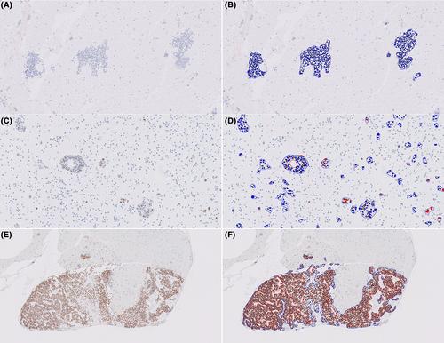
The study cohort comprised 105 consecutive cytology specimens with metastatic breast carcinoma. ER IHC WSIs were seamlessly integrated into the Visiopharm platform from the Image Management System (IMS) during our routine digital workflow, and an AI algorithm was employed for analysis. ER AI scores were compared with pathologists' manual consensus scores. Optimization steps were implemented and evaluated to reduce discordance.
Results
The overall concordance between pathologists' scores and AI scores was excellent (99/105, 94.3%). Six cases exhibited discordant results, including two false-negative (FN) cases due to abundant histiocytes incorrectly counted as negatively stained tumour cells by AI, two FN cases owing to weak staining, and two false-positive (FP) cases where pigmented macrophages were erroneously counted as positively stained tumour cells by AI. The Pearson correlation coefficient of ER-positive percentages between pathologists' and AI scores was 0.8483. Optimization steps, such as lowering the cut-off threshold and additional training using higher input magnification, significantly improved accuracy.
Conclusions
The automated ER AI algorithm demonstrated excellent concordance with pathologists' assessments and accurately differentiated ER-positive from ER-negative metastatic breast carcinoma cytology cases. However, precision in identifying tumour cells in cytology specimens requires further enhancement.
期刊介绍:
The aim of Cytopathology is to publish articles relating to those aspects of cytology which will increase our knowledge and understanding of the aetiology, diagnosis and management of human disease. It contains original articles and critical reviews on all aspects of clinical cytology in its broadest sense, including: gynaecological and non-gynaecological cytology; fine needle aspiration and screening strategy.
Cytopathology welcomes papers and articles on: ultrastructural, histochemical and immunocytochemical studies of the cell; quantitative cytology and DNA hybridization as applied to cytological material.

 求助内容:
求助内容: 应助结果提醒方式:


