Mohammadreza Dastouri, Hilal Ozdag, Ahmet Ruchan Akar, Alp Can
{"title":"诱导多能干细胞诱导内皮祖细胞和血管平滑肌细胞的分化及分子特性研究。","authors":"Mohammadreza Dastouri, Hilal Ozdag, Ahmet Ruchan Akar, Alp Can","doi":"10.34172/bi.2022.24132","DOIUrl":null,"url":null,"abstract":"<p><p></p><p><strong>Introduction: </strong>Pluripotent stem cells have been used by various researchers to differentiate and characterize endothelial cells (ECs) and vascular smooth muscle cells (VSMCs) for the clinical treatment of vascular injuries. Studies continue to differentiate and characterize the cells with higher vascularization potential and low risk of malignant transformation to the recipient. Unlike previous studies, this research aimed to differentiate induced pluripotent stem (iPS) cells into endothelial progenitor cells (EPCs) and VSMCs using a step-wise technique. This was achieved by elucidating the spatio-temporal expressions of the stage-specific genes and proteins during the differentiation process. The presence of highly expressed oncogenes in iPS cells was also investigated during the differentiation period.</p><p><strong>Methods: </strong>Induced PS cells were differentiated into lateral mesoderm cells (Flk1<sup>+</sup>). The Flk1<sup>+</sup> populations were isolated on day 5.5 of the mesodermal differentiation period. Flk1<sup>+</sup> cells were further differentiated into EPCs and VSMCs using VEGF<sub>165</sub> and platelet-derived growth factor-BB (PDGF-BB), respectively, and then characterized using gene expression levels, immunocytochemistry (ICC), and western blot (WB) methods. During the differentiation steps, the expression levels of the marker genes and proto-oncogenic <i>Myc</i> and <i>Klf4</i> genes were simultaneously studied.</p><p><strong>Results: </strong>The optimal time for the isolation of Flk1<sup>+</sup> cells was on day 5.5. EPCs and VSMCs were differentiated from Flk1<sup>+</sup> cells and characterized with EPC-specific markers, including <i>Kdr, Pecam1, CD133, Cdh5, Efnb2, Vcam1</i>; and VSMC-specific markers, including <i>Acta2, Cnn1, Des,</i> and <i>Myh11</i>. Differentiated cells were validated based on their temporal gene expressions, protein synthesis, and localization at certain time points. Significant decreases in <i>Myc</i> and <i>Klf4</i> gene expression levels were observed during the EPCs and VSMC differentiation period.</p><p><strong>Conclusion: </strong>EPCs and VSMCs were successfully differentiated from iPS cells and characterized by gene expression levels, ICC, and WB. We observed significant decreases in oncogene expression levels in the differentiated EPCs and VSMCs. In terms of safety, the described methodology provided a better safety margin. EPCs and VSMC obtained using this method may be good candidates for transplantation and vascular regeneration.</p>","PeriodicalId":48614,"journal":{"name":"Bioimpacts","volume":"13 4","pages":"289-300"},"PeriodicalIF":2.2000,"publicationDate":"2023-01-01","publicationTypes":"Journal Article","fieldsOfStudy":null,"isOpenAccess":false,"openAccessPdf":"https://ftp.ncbi.nlm.nih.gov/pub/pmc/oa_pdf/ae/cd/bi-13-289.PMC10460769.pdf","citationCount":"0","resultStr":"{\"title\":\"Differentiation and molecular characterization of endothelial progenitor and vascular smooth muscle cells from induced pluripotent stem cells.\",\"authors\":\"Mohammadreza Dastouri, Hilal Ozdag, Ahmet Ruchan Akar, Alp Can\",\"doi\":\"10.34172/bi.2022.24132\",\"DOIUrl\":null,\"url\":null,\"abstract\":\"<p><p></p><p><strong>Introduction: </strong>Pluripotent stem cells have been used by various researchers to differentiate and characterize endothelial cells (ECs) and vascular smooth muscle cells (VSMCs) for the clinical treatment of vascular injuries. Studies continue to differentiate and characterize the cells with higher vascularization potential and low risk of malignant transformation to the recipient. Unlike previous studies, this research aimed to differentiate induced pluripotent stem (iPS) cells into endothelial progenitor cells (EPCs) and VSMCs using a step-wise technique. This was achieved by elucidating the spatio-temporal expressions of the stage-specific genes and proteins during the differentiation process. The presence of highly expressed oncogenes in iPS cells was also investigated during the differentiation period.</p><p><strong>Methods: </strong>Induced PS cells were differentiated into lateral mesoderm cells (Flk1<sup>+</sup>). The Flk1<sup>+</sup> populations were isolated on day 5.5 of the mesodermal differentiation period. Flk1<sup>+</sup> cells were further differentiated into EPCs and VSMCs using VEGF<sub>165</sub> and platelet-derived growth factor-BB (PDGF-BB), respectively, and then characterized using gene expression levels, immunocytochemistry (ICC), and western blot (WB) methods. During the differentiation steps, the expression levels of the marker genes and proto-oncogenic <i>Myc</i> and <i>Klf4</i> genes were simultaneously studied.</p><p><strong>Results: </strong>The optimal time for the isolation of Flk1<sup>+</sup> cells was on day 5.5. EPCs and VSMCs were differentiated from Flk1<sup>+</sup> cells and characterized with EPC-specific markers, including <i>Kdr, Pecam1, CD133, Cdh5, Efnb2, Vcam1</i>; and VSMC-specific markers, including <i>Acta2, Cnn1, Des,</i> and <i>Myh11</i>. Differentiated cells were validated based on their temporal gene expressions, protein synthesis, and localization at certain time points. Significant decreases in <i>Myc</i> and <i>Klf4</i> gene expression levels were observed during the EPCs and VSMC differentiation period.</p><p><strong>Conclusion: </strong>EPCs and VSMCs were successfully differentiated from iPS cells and characterized by gene expression levels, ICC, and WB. We observed significant decreases in oncogene expression levels in the differentiated EPCs and VSMCs. In terms of safety, the described methodology provided a better safety margin. EPCs and VSMC obtained using this method may be good candidates for transplantation and vascular regeneration.</p>\",\"PeriodicalId\":48614,\"journal\":{\"name\":\"Bioimpacts\",\"volume\":\"13 4\",\"pages\":\"289-300\"},\"PeriodicalIF\":2.2000,\"publicationDate\":\"2023-01-01\",\"publicationTypes\":\"Journal Article\",\"fieldsOfStudy\":null,\"isOpenAccess\":false,\"openAccessPdf\":\"https://ftp.ncbi.nlm.nih.gov/pub/pmc/oa_pdf/ae/cd/bi-13-289.PMC10460769.pdf\",\"citationCount\":\"0\",\"resultStr\":null,\"platform\":\"Semanticscholar\",\"paperid\":null,\"PeriodicalName\":\"Bioimpacts\",\"FirstCategoryId\":\"5\",\"ListUrlMain\":\"https://doi.org/10.34172/bi.2022.24132\",\"RegionNum\":4,\"RegionCategory\":\"工程技术\",\"ArticlePicture\":[],\"TitleCN\":null,\"AbstractTextCN\":null,\"PMCID\":null,\"EPubDate\":\"\",\"PubModel\":\"\",\"JCR\":\"Q3\",\"JCRName\":\"PHARMACOLOGY & PHARMACY\",\"Score\":null,\"Total\":0}","platform":"Semanticscholar","paperid":null,"PeriodicalName":"Bioimpacts","FirstCategoryId":"5","ListUrlMain":"https://doi.org/10.34172/bi.2022.24132","RegionNum":4,"RegionCategory":"工程技术","ArticlePicture":[],"TitleCN":null,"AbstractTextCN":null,"PMCID":null,"EPubDate":"","PubModel":"","JCR":"Q3","JCRName":"PHARMACOLOGY & PHARMACY","Score":null,"Total":0}
引用次数: 0
摘要
多能干细胞已被许多研究人员用于分化和表征内皮细胞(ECs)和血管平滑肌细胞(VSMCs),用于血管损伤的临床治疗。研究继续分化和表征具有较高血管化潜力和对受体恶性转化风险低的细胞。与以往的研究不同,本研究旨在利用分步技术将诱导多能干细胞(iPS)分化为内皮祖细胞(EPCs)和VSMCs。这是通过阐明分化过程中阶段特异性基因和蛋白质的时空表达来实现的。在分化期间,还研究了iPS细胞中高表达癌基因的存在。方法:诱导PS细胞分化为外侧中胚层细胞(Flk1+)。Flk1+群体在中胚层分化期第5.5天分离。使用VEGF165和血小板衍生生长因子- bb (PDGF-BB)进一步将Flk1+细胞分化为EPCs和VSMCs,然后使用基因表达水平、免疫细胞化学(ICC)和western blot (WB)方法对其进行表征。在分化过程中,同时研究标记基因和原致癌基因Myc和Klf4的表达水平。结果:Flk1+细胞的最佳分离时间为第5.5天。从Flk1+细胞中分化出EPCs和VSMCs,并以EPCs特异性标记物Kdr、Pecam1、CD133、Cdh5、Efnb2、Vcam1进行表征;以及vsmc特异性标记物,包括Acta2、Cnn1、Des和Myh11。根据时间点的基因表达、蛋白质合成和定位来验证已分化的细胞。在EPCs和VSMC分化期间,Myc和Klf4基因表达水平显著降低。结论:从iPS细胞中成功分化出EPCs和VSMCs,并具有基因表达水平、ICC和WB的特征。我们观察到分化的EPCs和VSMCs中癌基因表达水平显著降低。在安全性方面,所描述的方法提供了更好的安全边际。用这种方法获得的内皮祖细胞和VSMC可能是移植和血管再生的良好候选者。
Differentiation and molecular characterization of endothelial progenitor and vascular smooth muscle cells from induced pluripotent stem cells.
Introduction: Pluripotent stem cells have been used by various researchers to differentiate and characterize endothelial cells (ECs) and vascular smooth muscle cells (VSMCs) for the clinical treatment of vascular injuries. Studies continue to differentiate and characterize the cells with higher vascularization potential and low risk of malignant transformation to the recipient. Unlike previous studies, this research aimed to differentiate induced pluripotent stem (iPS) cells into endothelial progenitor cells (EPCs) and VSMCs using a step-wise technique. This was achieved by elucidating the spatio-temporal expressions of the stage-specific genes and proteins during the differentiation process. The presence of highly expressed oncogenes in iPS cells was also investigated during the differentiation period.
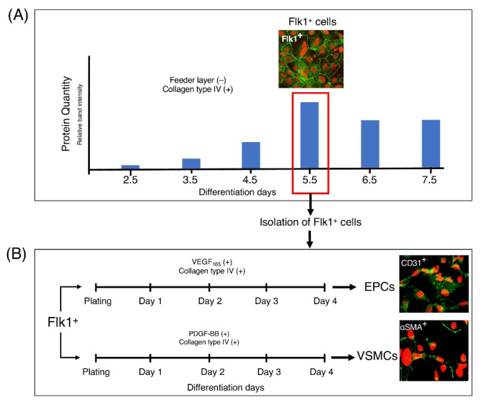
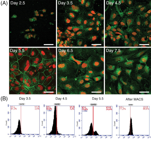
Methods: Induced PS cells were differentiated into lateral mesoderm cells (Flk1+). The Flk1+ populations were isolated on day 5.5 of the mesodermal differentiation period. Flk1+ cells were further differentiated into EPCs and VSMCs using VEGF165 and platelet-derived growth factor-BB (PDGF-BB), respectively, and then characterized using gene expression levels, immunocytochemistry (ICC), and western blot (WB) methods. During the differentiation steps, the expression levels of the marker genes and proto-oncogenic Myc and Klf4 genes were simultaneously studied.
Results: The optimal time for the isolation of Flk1+ cells was on day 5.5. EPCs and VSMCs were differentiated from Flk1+ cells and characterized with EPC-specific markers, including Kdr, Pecam1, CD133, Cdh5, Efnb2, Vcam1; and VSMC-specific markers, including Acta2, Cnn1, Des, and Myh11. Differentiated cells were validated based on their temporal gene expressions, protein synthesis, and localization at certain time points. Significant decreases in Myc and Klf4 gene expression levels were observed during the EPCs and VSMC differentiation period.
Conclusion: EPCs and VSMCs were successfully differentiated from iPS cells and characterized by gene expression levels, ICC, and WB. We observed significant decreases in oncogene expression levels in the differentiated EPCs and VSMCs. In terms of safety, the described methodology provided a better safety margin. EPCs and VSMC obtained using this method may be good candidates for transplantation and vascular regeneration.
BioimpactsPharmacology, Toxicology and Pharmaceutics-Pharmaceutical Science
CiteScore
4.80
自引率
7.70%
发文量
36
审稿时长
5 weeks
期刊介绍:
BioImpacts (BI) is a peer-reviewed multidisciplinary international journal, covering original research articles, reviews, commentaries, hypotheses, methodologies, and visions/reflections dealing with all aspects of biological and biomedical researches at molecular, cellular, functional and translational dimensions.

 求助内容:
求助内容: 应助结果提醒方式:


