{"title":"先天性心脏病患儿肺动脉的光学相干断层扫描:系统综述。","authors":"Ling Sun, Qiuping Jiang, Yumei Xie, Shushui Wang, Zhiwei Zhang","doi":"10.1002/ped4.12353","DOIUrl":null,"url":null,"abstract":"<p><strong>Importance: </strong>Optical coherence tomography (OCT) is a high-resolution intravascular imaging tool and has shown promise for providing real-time quantitative and qualitative descriptions of pulmonary vascular structures <i>in vivo</i> in adult pulmonary hypertension (PH), while not popular in pediatric patients with congenital heart diseases (CHD).</p><p><strong>Objective: </strong>The aim of this review is to summarize all the available evidence on the use of OCT for imaging pulmonary vascular remodeling in pediatric patients.</p><p><strong>Methods: </strong>We conducted the systematic literature resources (Cochran Library database, Medline via PubMed, EMBASE, and Web of Knowledge) from January 2010 to December 2021 and the search terms were \"PH\", \"child\", \"children\", \"pediatric\", \"OCT\", \"CHD\", \"pulmonary vessels\", \"pulmonary artery wall\". Studies in which OCT was used to image the pulmonary vessels in pediatric patients with CHD were considered for inclusion.</p><p><strong>Results: </strong>Five studies met the inclusion criteria. These five papers discussed the study of OCT in the pulmonary vasculature of different types of CHD, including common simple CHD, complex cyanotic CHD, and Williams-Beuren syndrome. In biventricular anatomy, pulmonary vascular remodeling was primarily reflected by pulmonary intima thickening from two-dimensional OCT. In single-ventricle anatomy, due to the state of hypoxia, the morphology of pulmonary vessels was indirectly reflected by the number and shape of nourishing vessels from three-dimensional OCT.</p><p><strong>Interpretation: </strong>OCT may be an adequate imaging procedure for the demonstration of pulmonary vascular structures and provide additional information in pediatric patients.</p>","PeriodicalId":19992,"journal":{"name":"Pediatric Investigation","volume":"6 4","pages":"264-270"},"PeriodicalIF":1.9000,"publicationDate":"2022-11-30","publicationTypes":"Journal Article","fieldsOfStudy":null,"isOpenAccess":false,"openAccessPdf":"https://ftp.ncbi.nlm.nih.gov/pub/pmc/oa_pdf/64/4a/PED4-6-264.PMC9789933.pdf","citationCount":"0","resultStr":"{\"title\":\"Optical coherence tomography of the pulmonary arteries in children with congenital heart diseases: A systematic review.\",\"authors\":\"Ling Sun, Qiuping Jiang, Yumei Xie, Shushui Wang, Zhiwei Zhang\",\"doi\":\"10.1002/ped4.12353\",\"DOIUrl\":null,\"url\":null,\"abstract\":\"<p><strong>Importance: </strong>Optical coherence tomography (OCT) is a high-resolution intravascular imaging tool and has shown promise for providing real-time quantitative and qualitative descriptions of pulmonary vascular structures <i>in vivo</i> in adult pulmonary hypertension (PH), while not popular in pediatric patients with congenital heart diseases (CHD).</p><p><strong>Objective: </strong>The aim of this review is to summarize all the available evidence on the use of OCT for imaging pulmonary vascular remodeling in pediatric patients.</p><p><strong>Methods: </strong>We conducted the systematic literature resources (Cochran Library database, Medline via PubMed, EMBASE, and Web of Knowledge) from January 2010 to December 2021 and the search terms were \\\"PH\\\", \\\"child\\\", \\\"children\\\", \\\"pediatric\\\", \\\"OCT\\\", \\\"CHD\\\", \\\"pulmonary vessels\\\", \\\"pulmonary artery wall\\\". Studies in which OCT was used to image the pulmonary vessels in pediatric patients with CHD were considered for inclusion.</p><p><strong>Results: </strong>Five studies met the inclusion criteria. These five papers discussed the study of OCT in the pulmonary vasculature of different types of CHD, including common simple CHD, complex cyanotic CHD, and Williams-Beuren syndrome. In biventricular anatomy, pulmonary vascular remodeling was primarily reflected by pulmonary intima thickening from two-dimensional OCT. In single-ventricle anatomy, due to the state of hypoxia, the morphology of pulmonary vessels was indirectly reflected by the number and shape of nourishing vessels from three-dimensional OCT.</p><p><strong>Interpretation: </strong>OCT may be an adequate imaging procedure for the demonstration of pulmonary vascular structures and provide additional information in pediatric patients.</p>\",\"PeriodicalId\":19992,\"journal\":{\"name\":\"Pediatric Investigation\",\"volume\":\"6 4\",\"pages\":\"264-270\"},\"PeriodicalIF\":1.9000,\"publicationDate\":\"2022-11-30\",\"publicationTypes\":\"Journal Article\",\"fieldsOfStudy\":null,\"isOpenAccess\":false,\"openAccessPdf\":\"https://ftp.ncbi.nlm.nih.gov/pub/pmc/oa_pdf/64/4a/PED4-6-264.PMC9789933.pdf\",\"citationCount\":\"0\",\"resultStr\":null,\"platform\":\"Semanticscholar\",\"paperid\":null,\"PeriodicalName\":\"Pediatric Investigation\",\"FirstCategoryId\":\"3\",\"ListUrlMain\":\"https://doi.org/10.1002/ped4.12353\",\"RegionNum\":4,\"RegionCategory\":\"医学\",\"ArticlePicture\":[],\"TitleCN\":null,\"AbstractTextCN\":null,\"PMCID\":null,\"EPubDate\":\"2022/12/1 0:00:00\",\"PubModel\":\"eCollection\",\"JCR\":\"Q2\",\"JCRName\":\"PEDIATRICS\",\"Score\":null,\"Total\":0}","platform":"Semanticscholar","paperid":null,"PeriodicalName":"Pediatric Investigation","FirstCategoryId":"3","ListUrlMain":"https://doi.org/10.1002/ped4.12353","RegionNum":4,"RegionCategory":"医学","ArticlePicture":[],"TitleCN":null,"AbstractTextCN":null,"PMCID":null,"EPubDate":"2022/12/1 0:00:00","PubModel":"eCollection","JCR":"Q2","JCRName":"PEDIATRICS","Score":null,"Total":0}
引用次数: 0
摘要
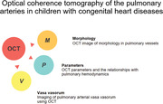
重要性:光学相干断层扫描(OCT)是一种高分辨率的血管内成像工具,可对成人肺动脉高压(PH)患者体内的肺血管结构进行实时定量和定性描述,但在患有先天性心脏病(CHD)的儿科患者中并不流行:本综述旨在总结有关使用 OCT 对儿科患者的肺血管重塑进行成像的所有现有证据:我们对 2010 年 1 月至 2021 年 12 月期间的系统性文献资源(Cochran 图书馆数据库、Medline via PubMed、EMBASE 和 Web of Knowledge)进行了检索,检索词为 "PH"、"儿童"、"儿童"、"儿科"、"OCT"、"CHD"、"肺血管"、"肺动脉壁"。结果显示,有五项研究符合纳入标准:结果:五项研究符合纳入标准。这五篇论文讨论了 OCT 对不同类型先天性心脏病(包括普通单纯先天性心脏病、复杂紫绀型先天性心脏病和 Williams-Beuren 综合征)肺血管的研究。在双心室解剖中,肺血管重塑主要通过二维 OCT 的肺内膜增厚来反映。在单心室解剖中,由于缺氧状态,肺血管的形态通过三维 OCT 的营养血管数量和形状间接反映出来:OCT可能是显示肺血管结构的适当成像程序,可为儿科患者提供更多信息。
Optical coherence tomography of the pulmonary arteries in children with congenital heart diseases: A systematic review.
Importance: Optical coherence tomography (OCT) is a high-resolution intravascular imaging tool and has shown promise for providing real-time quantitative and qualitative descriptions of pulmonary vascular structures in vivo in adult pulmonary hypertension (PH), while not popular in pediatric patients with congenital heart diseases (CHD).
Objective: The aim of this review is to summarize all the available evidence on the use of OCT for imaging pulmonary vascular remodeling in pediatric patients.
Methods: We conducted the systematic literature resources (Cochran Library database, Medline via PubMed, EMBASE, and Web of Knowledge) from January 2010 to December 2021 and the search terms were "PH", "child", "children", "pediatric", "OCT", "CHD", "pulmonary vessels", "pulmonary artery wall". Studies in which OCT was used to image the pulmonary vessels in pediatric patients with CHD were considered for inclusion.
Results: Five studies met the inclusion criteria. These five papers discussed the study of OCT in the pulmonary vasculature of different types of CHD, including common simple CHD, complex cyanotic CHD, and Williams-Beuren syndrome. In biventricular anatomy, pulmonary vascular remodeling was primarily reflected by pulmonary intima thickening from two-dimensional OCT. In single-ventricle anatomy, due to the state of hypoxia, the morphology of pulmonary vessels was indirectly reflected by the number and shape of nourishing vessels from three-dimensional OCT.
Interpretation: OCT may be an adequate imaging procedure for the demonstration of pulmonary vascular structures and provide additional information in pediatric patients.
 求助内容:
求助内容: 应助结果提醒方式:


