T Michael Nork, Charlene B Y Kim, Alexander W Katz, Carol A Rasmussen, Mark Banghart, James N Ver Hoeve
{"title":"Multifocal electroretinography increases following experimental glaucoma in nonhuman primates with retinal ganglion cell axotomy.","authors":"T Michael Nork, Charlene B Y Kim, Alexander W Katz, Carol A Rasmussen, Mark Banghart, James N Ver Hoeve","doi":"10.1007/s10633-023-09922-1","DOIUrl":null,"url":null,"abstract":"<p><strong>Purpose: </strong>To determine whether short-latency changes in multifocal electroretinography (mfERG) observed in experimental glaucoma (EG) are secondary solely to retinal ganglion cell (RGC) loss or whether there is a separate contribution from elevated intraocular pressure (IOP).</p><p><strong>Methods: </strong>Prior to operative procedures, a series of baseline mfERGs were recorded from six rhesus macaques using a 241-element unstretched stimulus. Animals then underwent hemiretinal endodiathermy axotomy (HEA) by placing burns along the inferior 180° of the optic nerve margin in the right eye (OD). mfERG recordings were obtained in each animal at regular intervals following for 3-4 months to allow stabilization of the HEA effects. Laser trabecular meshwork destruction (LTD) to elevate IOP was then performed; first-order kernel (K1) waveform root-mean-square (RMS) amplitudes for the short-latency segment of the mfERG wave (9-35 ms) were computed for two 7-hexagon groupings-the first located within the superior (non-axotomized) macula and the second within the inferior (axotomized) macula. Immunohistochemistry for glial fibrillary acidic protein (GFAP) was done.</p><p><strong>Results: </strong>By 3 months post HEA, there was marked thinning of the inferior nerve fiber layer as measured by optical coherence tomography. Compared with baseline, no statistically significant changes in 9-35 ms K1 RMS amplitudes were evident in either the axotomized or non-axotomized portions of the macula. Following LTD, mean IOP in HEA eyes rose to 46 ± 9 compared with 20 ± 2 mmHg (SD) in the fellow control eyes. In the HEA + EG eyes, statistically significant increases in K1 RMS amplitude were present in both the axotomized inferior and non-axotomized superior portions of the OD retinas. No changes in K1 RMS amplitude were found in the fellow control eyes from baseline to HEA epoch, but there was a smaller increase from baseline to HEA + EG. Upregulation of GFAP in the Müller cells was evident in both non-axotomized and axotomized retina in eyes with elevated IOP.</p><p><strong>Conclusions: </strong>The RMS amplitudes of the short-latency mfERG K1 waveforms are not altered following axotomy but undergo marked increases following elevated IOP. This suggests that the increase in mfERG amplitude was not solely a result of RGC loss and may reflect photoreceptor and bipolar cell dysfunction and/or changes in Müller cells.</p>","PeriodicalId":11207,"journal":{"name":"Documenta Ophthalmologica","volume":"146 2","pages":"97-112"},"PeriodicalIF":2.6000,"publicationDate":"2023-04-01","publicationTypes":"Journal Article","fieldsOfStudy":null,"isOpenAccess":false,"openAccessPdf":"https://www.ncbi.nlm.nih.gov/pmc/articles/PMC10284020/pdf/","citationCount":"0","resultStr":null,"platform":"Semanticscholar","paperid":null,"PeriodicalName":"Documenta Ophthalmologica","FirstCategoryId":"3","ListUrlMain":"https://doi.org/10.1007/s10633-023-09922-1","RegionNum":4,"RegionCategory":"医学","ArticlePicture":[],"TitleCN":null,"AbstractTextCN":null,"PMCID":null,"EPubDate":"2023/2/10 0:00:00","PubModel":"Epub","JCR":"Q2","JCRName":"OPHTHALMOLOGY","Score":null,"Total":0}
引用次数: 0
Abstract
Purpose: To determine whether short-latency changes in multifocal electroretinography (mfERG) observed in experimental glaucoma (EG) are secondary solely to retinal ganglion cell (RGC) loss or whether there is a separate contribution from elevated intraocular pressure (IOP).
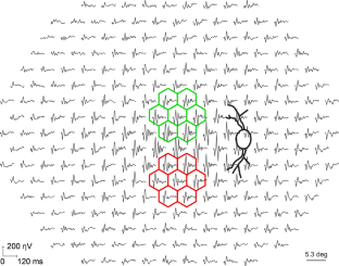
Methods: Prior to operative procedures, a series of baseline mfERGs were recorded from six rhesus macaques using a 241-element unstretched stimulus. Animals then underwent hemiretinal endodiathermy axotomy (HEA) by placing burns along the inferior 180° of the optic nerve margin in the right eye (OD). mfERG recordings were obtained in each animal at regular intervals following for 3-4 months to allow stabilization of the HEA effects. Laser trabecular meshwork destruction (LTD) to elevate IOP was then performed; first-order kernel (K1) waveform root-mean-square (RMS) amplitudes for the short-latency segment of the mfERG wave (9-35 ms) were computed for two 7-hexagon groupings-the first located within the superior (non-axotomized) macula and the second within the inferior (axotomized) macula. Immunohistochemistry for glial fibrillary acidic protein (GFAP) was done.
Results: By 3 months post HEA, there was marked thinning of the inferior nerve fiber layer as measured by optical coherence tomography. Compared with baseline, no statistically significant changes in 9-35 ms K1 RMS amplitudes were evident in either the axotomized or non-axotomized portions of the macula. Following LTD, mean IOP in HEA eyes rose to 46 ± 9 compared with 20 ± 2 mmHg (SD) in the fellow control eyes. In the HEA + EG eyes, statistically significant increases in K1 RMS amplitude were present in both the axotomized inferior and non-axotomized superior portions of the OD retinas. No changes in K1 RMS amplitude were found in the fellow control eyes from baseline to HEA epoch, but there was a smaller increase from baseline to HEA + EG. Upregulation of GFAP in the Müller cells was evident in both non-axotomized and axotomized retina in eyes with elevated IOP.
Conclusions: The RMS amplitudes of the short-latency mfERG K1 waveforms are not altered following axotomy but undergo marked increases following elevated IOP. This suggests that the increase in mfERG amplitude was not solely a result of RGC loss and may reflect photoreceptor and bipolar cell dysfunction and/or changes in Müller cells.
期刊介绍:
Documenta Ophthalmologica is an official publication of the International Society for Clinical Electrophysiology of Vision. The purpose of the journal is to promote the understanding and application of clinical electrophysiology of vision. Documenta Ophthalmologica will publish reviews, research articles, technical notes, brief reports and case studies which inform the readers about basic and clinical sciences related to visual electrodiagnosis and means to improve diagnosis and clinical management of patients using visual electrophysiology. Studies may involve animals or humans. In either case appropriate care must be taken to follow the Declaration of Helsinki for human subject or appropriate humane standards of animal care (e.g., the ARVO standards on Animal Care and Use).

 求助内容:
求助内容: 应助结果提醒方式:


