{"title":"Electroretinographic abnormalities in Alport syndrome with a novel COL4A5 truncated variant (p.Try20GlyfsTer19).","authors":"Kei Mizobuchi, Takaaki Hayashi, Ryo Ohira, Tadashi Nakano","doi":"10.1007/s10633-023-09935-w","DOIUrl":null,"url":null,"abstract":"<p><strong>Purpose: </strong>Alport syndrome comprises a heterogeneous group of inherited kidney diseases that are associated with ocular complications. In this study, we aimed to detail the clinical characteristics of a patient with X-linked Alport syndrome.</p><p><strong>Methods: </strong>We performed next-generation sequencing (NGS) with hybridization capture to identify the disease-causing variant of Alport syndrome and a comprehensive ophthalmic examination, including full-field electroretinography (FF-ERG).</p><p><strong>Results: </strong>Genetic testing using NGS with hybridization capture revealed a novel hemizygous variant [c.51_52delGA (p.Trp20GlyfsTer19)] in exon 1 of COL4A5. The patient underwent cataract surgery in both eyes because of decreased visual acuity and photophobia. The best-corrected visual acuity improved from 0.9 and 0.7 in the right and left eyes, respectively, to 1.5 in both eyes. Anterior-segment optical coherence tomography (OCT) revealed anterior and posterior lenticonus. Fundus photographs showed central and peripheral fleck retinopathy. Wide-field fundus autofluorescence (AF) imaging showed mottled hyper- and hypo-AF in the peripheral retina, which was consistent with peripheral fleck retinopathy. Furthermore, OCT revealed thinning of the inner retinal layers, especially at the temporal macular, but the outer retinal layers were preserved. Ganglion cell analysis showed no progression for 5 years. FF-ERG was performed at 41 (phakia) and 46 (pseudophakia) years of age. The amplitudes of dark-adapted (DA) and light-adapted (LA) responses showed selective b-wave abnormalities. The b/a-wave ratios of DA 3.0 were 1.22 and 1.16 in the right and left eyes, respectively. The amplitudes of DA 3.0 oscillatory potentials (OP) were reduced. Five years later, the amplitudes of DA and LA responses revealed no remarkable changes, except for an OP wave of DA 3.0, which was substantially reduced.</p><p><strong>Conclusions: </strong>Our findings revealed electroretinographic abnormalities in a patient with Alport syndrome, which predominantly indicated impairment of the inner retina. Notably, little short-term progression was observed.</p>","PeriodicalId":11207,"journal":{"name":"Documenta Ophthalmologica","volume":"146 3","pages":"281-291"},"PeriodicalIF":2.9000,"publicationDate":"2023-06-01","publicationTypes":"Journal Article","fieldsOfStudy":null,"isOpenAccess":false,"openAccessPdf":"","citationCount":"0","resultStr":null,"platform":"Semanticscholar","paperid":null,"PeriodicalName":"Documenta Ophthalmologica","FirstCategoryId":"3","ListUrlMain":"https://doi.org/10.1007/s10633-023-09935-w","RegionNum":4,"RegionCategory":"医学","ArticlePicture":[],"TitleCN":null,"AbstractTextCN":null,"PMCID":null,"EPubDate":"2023/5/10 0:00:00","PubModel":"Epub","JCR":"Q2","JCRName":"OPHTHALMOLOGY","Score":null,"Total":0}
引用次数: 0
Abstract
Purpose: Alport syndrome comprises a heterogeneous group of inherited kidney diseases that are associated with ocular complications. In this study, we aimed to detail the clinical characteristics of a patient with X-linked Alport syndrome.
Methods: We performed next-generation sequencing (NGS) with hybridization capture to identify the disease-causing variant of Alport syndrome and a comprehensive ophthalmic examination, including full-field electroretinography (FF-ERG).
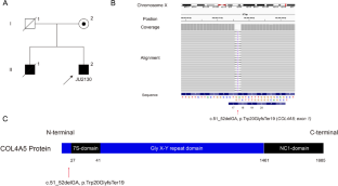
Results: Genetic testing using NGS with hybridization capture revealed a novel hemizygous variant [c.51_52delGA (p.Trp20GlyfsTer19)] in exon 1 of COL4A5. The patient underwent cataract surgery in both eyes because of decreased visual acuity and photophobia. The best-corrected visual acuity improved from 0.9 and 0.7 in the right and left eyes, respectively, to 1.5 in both eyes. Anterior-segment optical coherence tomography (OCT) revealed anterior and posterior lenticonus. Fundus photographs showed central and peripheral fleck retinopathy. Wide-field fundus autofluorescence (AF) imaging showed mottled hyper- and hypo-AF in the peripheral retina, which was consistent with peripheral fleck retinopathy. Furthermore, OCT revealed thinning of the inner retinal layers, especially at the temporal macular, but the outer retinal layers were preserved. Ganglion cell analysis showed no progression for 5 years. FF-ERG was performed at 41 (phakia) and 46 (pseudophakia) years of age. The amplitudes of dark-adapted (DA) and light-adapted (LA) responses showed selective b-wave abnormalities. The b/a-wave ratios of DA 3.0 were 1.22 and 1.16 in the right and left eyes, respectively. The amplitudes of DA 3.0 oscillatory potentials (OP) were reduced. Five years later, the amplitudes of DA and LA responses revealed no remarkable changes, except for an OP wave of DA 3.0, which was substantially reduced.
Conclusions: Our findings revealed electroretinographic abnormalities in a patient with Alport syndrome, which predominantly indicated impairment of the inner retina. Notably, little short-term progression was observed.
期刊介绍:
Documenta Ophthalmologica is an official publication of the International Society for Clinical Electrophysiology of Vision. The purpose of the journal is to promote the understanding and application of clinical electrophysiology of vision. Documenta Ophthalmologica will publish reviews, research articles, technical notes, brief reports and case studies which inform the readers about basic and clinical sciences related to visual electrodiagnosis and means to improve diagnosis and clinical management of patients using visual electrophysiology. Studies may involve animals or humans. In either case appropriate care must be taken to follow the Declaration of Helsinki for human subject or appropriate humane standards of animal care (e.g., the ARVO standards on Animal Care and Use).

 求助内容:
求助内容: 应助结果提醒方式:


