{"title":"Brain diffusion tensor imaging reveals altered connections and networks in epilepsy patients.","authors":"Meixia Wang, Xiaoyu Cheng, Qianru Shi, Bo Xu, Xiaoxia Hou, Huimin Zhao, Qian Gui, Guanhui Wu, Xiaofeng Dong, Qinrong Xu, Mingqiang Shen, Qingzhang Cheng, Shouru Xue, Hongxuan Feng, Zhiliang Ding","doi":"10.3389/fnhum.2023.1142408","DOIUrl":null,"url":null,"abstract":"<p><strong>Introduction: </strong>Accumulating evidence shows that epilepsy is a disease caused by brain network dysfunction. This study explored changes in brain network structure in epilepsy patients based on graph analysis of diffusion tensor imaging data.</p><p><strong>Methods: </strong>The brain structure networks of 42 healthy control individuals and 26 epilepsy patients were constructed. Using graph theory analysis, global and local network topology parameters of the brain structure network were calculated, and changes in global and local characteristics of the brain network in epilepsy patients were quantitatively analyzed.</p><p><strong>Results: </strong>Compared with the healthy control group, the epilepsy patient group showed lower global efficiency, local efficiency, clustering coefficient, and a longer shortest path length. Both healthy control individuals and epilepsy patients showed small-world attributes, with no significant difference between groups. The epilepsy patient group showed lower nodal local efficiency and nodal clustering coefficient in the right olfactory cortex and right rectus and lower nodal degree centrality in the right olfactory cortex and the left paracentral lobular compared with the healthy control group. In addition, the epilepsy patient group showed a smaller fiber number of edges in specific regions of the frontal lobe, temporal lobe, and default mode network, indicating reduced connection strength.</p><p><strong>Discussion: </strong>Epilepsy patients exhibited lower global and local brain network properties as well as reduced white matter fiber connectivity in key brain regions. These findings further support the idea that epilepsy is a brain network disorder.</p>","PeriodicalId":12536,"journal":{"name":"Frontiers in Human Neuroscience","volume":"17 ","pages":"1142408"},"PeriodicalIF":2.4000,"publicationDate":"2023-01-01","publicationTypes":"Journal Article","fieldsOfStudy":null,"isOpenAccess":false,"openAccessPdf":"https://www.ncbi.nlm.nih.gov/pmc/articles/PMC10073437/pdf/","citationCount":"1","resultStr":null,"platform":"Semanticscholar","paperid":null,"PeriodicalName":"Frontiers in Human Neuroscience","FirstCategoryId":"3","ListUrlMain":"https://doi.org/10.3389/fnhum.2023.1142408","RegionNum":3,"RegionCategory":"医学","ArticlePicture":[],"TitleCN":null,"AbstractTextCN":null,"PMCID":null,"EPubDate":"","PubModel":"","JCR":"Q3","JCRName":"NEUROSCIENCES","Score":null,"Total":0}
引用次数: 1
Abstract
Introduction: Accumulating evidence shows that epilepsy is a disease caused by brain network dysfunction. This study explored changes in brain network structure in epilepsy patients based on graph analysis of diffusion tensor imaging data.
Methods: The brain structure networks of 42 healthy control individuals and 26 epilepsy patients were constructed. Using graph theory analysis, global and local network topology parameters of the brain structure network were calculated, and changes in global and local characteristics of the brain network in epilepsy patients were quantitatively analyzed.
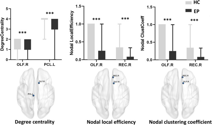
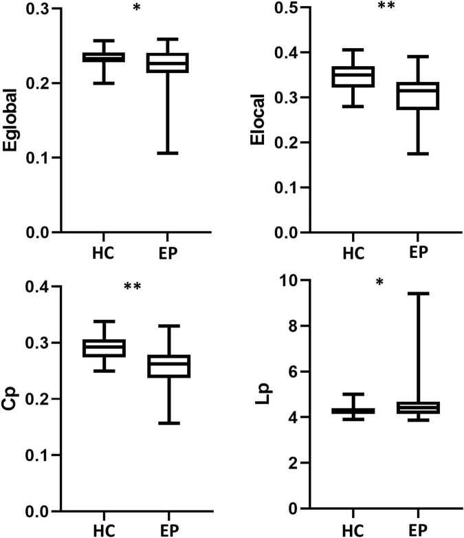
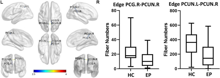
Results: Compared with the healthy control group, the epilepsy patient group showed lower global efficiency, local efficiency, clustering coefficient, and a longer shortest path length. Both healthy control individuals and epilepsy patients showed small-world attributes, with no significant difference between groups. The epilepsy patient group showed lower nodal local efficiency and nodal clustering coefficient in the right olfactory cortex and right rectus and lower nodal degree centrality in the right olfactory cortex and the left paracentral lobular compared with the healthy control group. In addition, the epilepsy patient group showed a smaller fiber number of edges in specific regions of the frontal lobe, temporal lobe, and default mode network, indicating reduced connection strength.
Discussion: Epilepsy patients exhibited lower global and local brain network properties as well as reduced white matter fiber connectivity in key brain regions. These findings further support the idea that epilepsy is a brain network disorder.
期刊介绍:
Frontiers in Human Neuroscience is a first-tier electronic journal devoted to understanding the brain mechanisms supporting cognitive and social behavior in humans, and how these mechanisms might be altered in disease states. The last 25 years have seen an explosive growth in both the methods and the theoretical constructs available to study the human brain. Advances in electrophysiological, neuroimaging, neuropsychological, psychophysical, neuropharmacological and computational approaches have provided key insights into the mechanisms of a broad range of human behaviors in both health and disease. Work in human neuroscience ranges from the cognitive domain, including areas such as memory, attention, language and perception to the social domain, with this last subject addressing topics, such as interpersonal interactions, social discourse and emotional regulation. How these processes unfold during development, mature in adulthood and often decline in aging, and how they are altered in a host of developmental, neurological and psychiatric disorders, has become increasingly amenable to human neuroscience research approaches. Work in human neuroscience has influenced many areas of inquiry ranging from social and cognitive psychology to economics, law and public policy. Accordingly, our journal will provide a forum for human research spanning all areas of human cognitive, social, developmental and translational neuroscience using any research approach.

 求助内容:
求助内容: 应助结果提醒方式:


