Austin Pereira, Birgit Ertl-Wagner, Anupreet Tumber, Ajoy Vincent, Michael J Wan
{"title":"Bilateral compressive optic neuropathy and outer retinopathy due to optic canal hyperostosis in a child with isolated vitamin a deficiency.","authors":"Austin Pereira, Birgit Ertl-Wagner, Anupreet Tumber, Ajoy Vincent, Michael J Wan","doi":"10.1007/s10633-022-09918-3","DOIUrl":null,"url":null,"abstract":"<p><strong>Purpose: </strong>Vitamin A plays a crucial role in rod phototransduction, with deficient levels manifesting as night blindness. Animal models have demonstrated bone dysplasia in the setting of hypovitaminosis A. We present a rare case of bony overgrowth leading to bilateral compressive optic neuropathy, combined with outer retinopathy, in a paediatric patient secondary to isolated vitamin A deficiency.</p><p><strong>Methods: </strong>A single case report was conducted from Toronto, Canada.</p><p><strong>Results: </strong>A 12-year-old boy with known autism spectrum disorder presented with a 9-month history of progressive painless vision loss. Vision was 20/300 and hand motion in the right and left eye, respectively. Fundus photography demonstrated bilateral optic atrophy and yellow lesions notably in the right eye far periphery. Optical coherence tomography (OCT) imaging demonstrated thinning of the retinal nerve fibre layer, alterations in the ellipsoid zone, as well as retinal pigment epithelium deposits. Computed tomography imaging demonstrated sphenoid bone thickening with narrow optic canals and moderate optic atrophy bilaterally. Full-field electroretinogram (ERG) demonstrated mildly reduced dark adapted (DA) 0.01 b-wave amplitudes and electronegative configuration of DA 3.0 and DA 10.0 ERG; the light adapted ERGs were normal. The patient was treated with pulse vitamin A therapy. Subsequently, the DA ERG normalized, outer retinal changes reversed and vision stabilised; no surgical intervention was conducted.</p><p><strong>Conclusion: </strong>This case represents a rare presentation of compressive optic neuropathy with concomitant outer retinopathy secondary to isolated vitamin A deficiency. Despite improvement in outer retinal integrity on OCT imaging and ERG testing results following vitamin A supplementation, no functional improvement was obtained due to severe optic atrophy.</p>","PeriodicalId":11207,"journal":{"name":"Documenta Ophthalmologica","volume":"146 2","pages":"173-180"},"PeriodicalIF":2.6000,"publicationDate":"2023-04-01","publicationTypes":"Journal Article","fieldsOfStudy":null,"isOpenAccess":false,"openAccessPdf":"","citationCount":"2","resultStr":null,"platform":"Semanticscholar","paperid":null,"PeriodicalName":"Documenta Ophthalmologica","FirstCategoryId":"3","ListUrlMain":"https://doi.org/10.1007/s10633-022-09918-3","RegionNum":4,"RegionCategory":"医学","ArticlePicture":[],"TitleCN":null,"AbstractTextCN":null,"PMCID":null,"EPubDate":"","PubModel":"","JCR":"Q2","JCRName":"OPHTHALMOLOGY","Score":null,"Total":0}
引用次数: 2
Abstract
Purpose: Vitamin A plays a crucial role in rod phototransduction, with deficient levels manifesting as night blindness. Animal models have demonstrated bone dysplasia in the setting of hypovitaminosis A. We present a rare case of bony overgrowth leading to bilateral compressive optic neuropathy, combined with outer retinopathy, in a paediatric patient secondary to isolated vitamin A deficiency.
Methods: A single case report was conducted from Toronto, Canada.
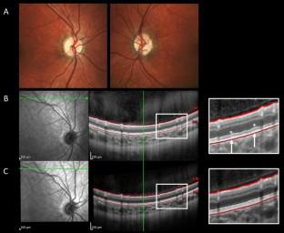
Results: A 12-year-old boy with known autism spectrum disorder presented with a 9-month history of progressive painless vision loss. Vision was 20/300 and hand motion in the right and left eye, respectively. Fundus photography demonstrated bilateral optic atrophy and yellow lesions notably in the right eye far periphery. Optical coherence tomography (OCT) imaging demonstrated thinning of the retinal nerve fibre layer, alterations in the ellipsoid zone, as well as retinal pigment epithelium deposits. Computed tomography imaging demonstrated sphenoid bone thickening with narrow optic canals and moderate optic atrophy bilaterally. Full-field electroretinogram (ERG) demonstrated mildly reduced dark adapted (DA) 0.01 b-wave amplitudes and electronegative configuration of DA 3.0 and DA 10.0 ERG; the light adapted ERGs were normal. The patient was treated with pulse vitamin A therapy. Subsequently, the DA ERG normalized, outer retinal changes reversed and vision stabilised; no surgical intervention was conducted.
Conclusion: This case represents a rare presentation of compressive optic neuropathy with concomitant outer retinopathy secondary to isolated vitamin A deficiency. Despite improvement in outer retinal integrity on OCT imaging and ERG testing results following vitamin A supplementation, no functional improvement was obtained due to severe optic atrophy.
期刊介绍:
Documenta Ophthalmologica is an official publication of the International Society for Clinical Electrophysiology of Vision. The purpose of the journal is to promote the understanding and application of clinical electrophysiology of vision. Documenta Ophthalmologica will publish reviews, research articles, technical notes, brief reports and case studies which inform the readers about basic and clinical sciences related to visual electrodiagnosis and means to improve diagnosis and clinical management of patients using visual electrophysiology. Studies may involve animals or humans. In either case appropriate care must be taken to follow the Declaration of Helsinki for human subject or appropriate humane standards of animal care (e.g., the ARVO standards on Animal Care and Use).

 求助内容:
求助内容: 应助结果提醒方式:


