Miniature microscopes for manipulating and recording in vivo brain activity
IF 1.8
4区 工程技术
引用次数: 15
Abstract
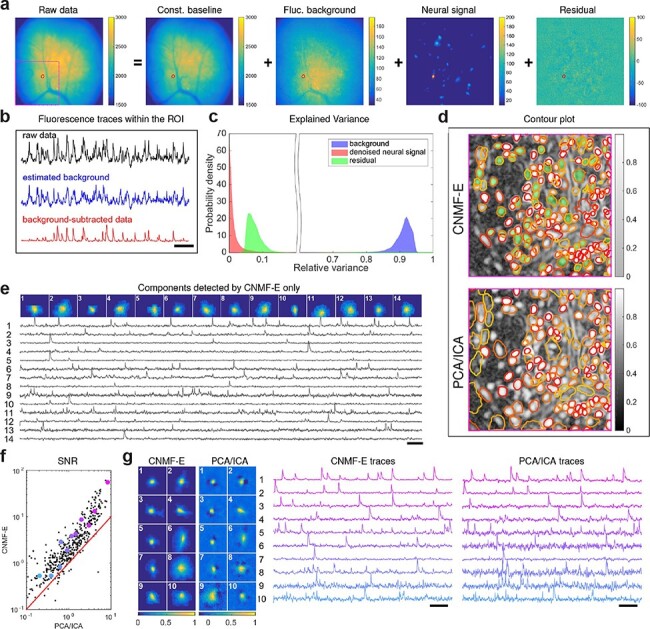
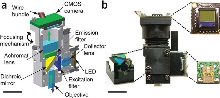
Here we describe the development and application of miniature integrated microscopes (miniscopes) paired with microendoscopes that allow for the visualization and manipulation of neural circuits in superficial and subcortical brain regions in freely behaving animals. Over the past decade the miniscope platform has expanded to include simultaneous optogenetic capabilities, electrically-tunable lenses that enable multi-plane imaging, color-corrected optics, and an integrated data acquisition platform that streamlines multimodal experiments. Miniscopes have given researchers an unprecedented ability to monitor hundreds to thousands of genetically-defined neurons from weeks to months in both healthy and diseased animal brains. Sophisticated algorithms that take advantage of constrained matrix factorization allow for background estimation and reliable cell identification, greatly improving the reliability and scalability of source extraction for large imaging datasets. Data generated from miniscopes have empowered researchers to investigate the neural circuit underpinnings of a wide array of behaviors that cannot be studied under head-fixed conditions, such as sleep, reward seeking, learning and memory, social behaviors, and feeding. Importantly, the miniscope has broadened our understanding of how neural circuits can go awry in animal models of progressive neurological disorders, such as Parkinson's disease. Continued miniscope development, including the ability to record from multiple populations of cells simultaneously, along with continued multimodal integration of techniques such as electrophysiology, will allow for deeper understanding into the neural circuits that underlie complex and naturalistic behavior.


用于操纵和记录体内大脑活动的微型显微镜
在这里,我们描述了微型集成显微镜(miniscope)与微型内窥镜的开发和应用,该显微镜可对行为自由的动物大脑浅表和皮层下区域的神经回路进行可视化和操作。在过去的十年里,迷你镜平台已经扩展到包括同时光遗传学功能、实现多平面成像的电可调谐透镜、颜色校正光学器件,以及简化多模式实验的集成数据采集平台。微型示波器为研究人员提供了前所未有的能力,可以在数周至数月内监测健康和患病动物大脑中数百至数千个基因定义的神经元。利用约束矩阵分解的复杂算法可以进行背景估计和可靠的细胞识别,大大提高了大型成像数据集源提取的可靠性和可扩展性。微型示波器产生的数据使研究人员能够研究一系列在固定头部条件下无法研究的行为的神经回路基础,如睡眠、寻求奖励、学习和记忆、社交行为和进食。重要的是,迷你显微镜拓宽了我们对神经回路如何在进展性神经疾病(如帕金森病)的动物模型中出错的理解。持续的微型示波器开发,包括同时记录多个细胞群的能力,以及电生理学等技术的持续多模式集成,将使人们能够更深入地了解复杂和自然行为背后的神经回路。
本文章由计算机程序翻译,如有差异,请以英文原文为准。
求助全文
约1分钟内获得全文
求助全文
来源期刊

Microscopy
工程技术-显微镜技术
自引率
11.10%
发文量
0
审稿时长
>12 weeks
期刊介绍:
Microscopy, previously Journal of Electron Microscopy, promotes research combined with any type of microscopy techniques, applied in life and material sciences. Microscopy is the official journal of the Japanese Society of Microscopy.
 求助内容:
求助内容: 应助结果提醒方式:


