Jinyue Dai, Xiaofei Wang, YingXiang Han, Cancan Xue, XuRan Dong, Ke Zhang, Haocheng Xian, WeiJia Zhang, Chun Zhang, Jing Hong
{"title":"High Myopia-Induced Optic Nerve Head Deformation and Glaucoma Progression: A Three-Year Follow-Up Study.","authors":"Jinyue Dai, Xiaofei Wang, YingXiang Han, Cancan Xue, XuRan Dong, Ke Zhang, Haocheng Xian, WeiJia Zhang, Chun Zhang, Jing Hong","doi":"10.1167/iovs.66.13.30","DOIUrl":null,"url":null,"abstract":"<p><strong>Purpose: </strong>To investigate the biomechanical effects of high myopia (HM)-related optic nerve head (ONH) structural changes on glaucoma progression and identify key predictive parameters.</p><p><strong>Methods: </strong>This prospective cohort study enrolled 242 eyes: 97 highly myopic glaucoma (HMG), axial length > 26.5 mm; 145 open-angle glaucoma (OAG), axial length ≤ 26.5 mm. ONH parameters, including Bruch's membrane opening (BMO), anterior scleral canal opening (ASCO) areas, neural canal minimum cross-sectional area (NCMCA), and ASCO-BMO offset, were quantified via spectral-domain OCT. Visual field (VF) defects and parapapillary retinal nerve fiber layer (pRNFL) thickness were monitored over 3 years. Generalized linear mixed models and multivariate regression were used for analysis.</p><p><strong>Results: </strong>Compared with OAG, HMG eyes exhibited larger BMO areas (3.0 vs. 2.3 mm2, P < 0.001) and ASCO areas (2.6 vs. 2.4 mm2, P < 0.001), greater ASCO-BMO offset (376.1 vs. 161.5 µm, P < 0.001), smaller NCMCA (1.0 vs. 1.3 mm2, P < 0.001), and faster temporal VF progression (0.173 vs. 0.060 dB/y, P = 0.023) and pRNFL thinning (0.96 vs. 0.64 µm/y, P = 0.014). Multivariate analysis identified NCMCA as an independent predictor of global progression (β = -0.03, P = 0.021).</p><p><strong>Conclusions: </strong>HM-induced ONH deformation, particularly nasal tilting and reduced NCMCA, accelerates temporal glaucomatous damage. NCMCA serves as a critical biomarker for progression risk, supporting individualized management in glaucoma with HM.</p>","PeriodicalId":14620,"journal":{"name":"Investigative ophthalmology & visual science","volume":"66 13","pages":"30"},"PeriodicalIF":4.7000,"publicationDate":"2025-10-01","publicationTypes":"Journal Article","fieldsOfStudy":null,"isOpenAccess":false,"openAccessPdf":"https://www.ncbi.nlm.nih.gov/pmc/articles/PMC12534888/pdf/","citationCount":"0","resultStr":null,"platform":"Semanticscholar","paperid":null,"PeriodicalName":"Investigative ophthalmology & visual science","FirstCategoryId":"3","ListUrlMain":"https://doi.org/10.1167/iovs.66.13.30","RegionNum":2,"RegionCategory":"医学","ArticlePicture":[],"TitleCN":null,"AbstractTextCN":null,"PMCID":null,"EPubDate":"","PubModel":"","JCR":"Q1","JCRName":"OPHTHALMOLOGY","Score":null,"Total":0}
引用次数: 0
Abstract
Purpose: To investigate the biomechanical effects of high myopia (HM)-related optic nerve head (ONH) structural changes on glaucoma progression and identify key predictive parameters.
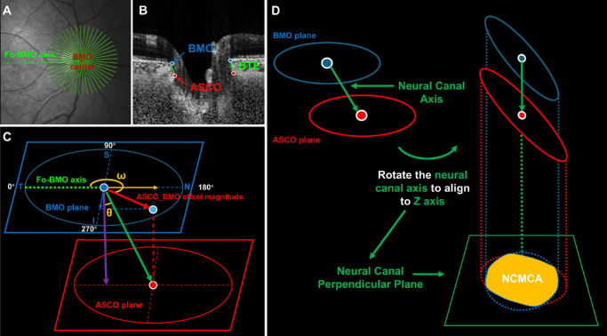
Methods: This prospective cohort study enrolled 242 eyes: 97 highly myopic glaucoma (HMG), axial length > 26.5 mm; 145 open-angle glaucoma (OAG), axial length ≤ 26.5 mm. ONH parameters, including Bruch's membrane opening (BMO), anterior scleral canal opening (ASCO) areas, neural canal minimum cross-sectional area (NCMCA), and ASCO-BMO offset, were quantified via spectral-domain OCT. Visual field (VF) defects and parapapillary retinal nerve fiber layer (pRNFL) thickness were monitored over 3 years. Generalized linear mixed models and multivariate regression were used for analysis.
Results: Compared with OAG, HMG eyes exhibited larger BMO areas (3.0 vs. 2.3 mm2, P < 0.001) and ASCO areas (2.6 vs. 2.4 mm2, P < 0.001), greater ASCO-BMO offset (376.1 vs. 161.5 µm, P < 0.001), smaller NCMCA (1.0 vs. 1.3 mm2, P < 0.001), and faster temporal VF progression (0.173 vs. 0.060 dB/y, P = 0.023) and pRNFL thinning (0.96 vs. 0.64 µm/y, P = 0.014). Multivariate analysis identified NCMCA as an independent predictor of global progression (β = -0.03, P = 0.021).
Conclusions: HM-induced ONH deformation, particularly nasal tilting and reduced NCMCA, accelerates temporal glaucomatous damage. NCMCA serves as a critical biomarker for progression risk, supporting individualized management in glaucoma with HM.
目的:探讨高度近视(HM)相关视神经头(ONH)结构改变对青光眼进展的生物力学影响,并确定关键预测参数。方法:本前瞻性队列研究纳入242只眼:高度近视青光眼(HMG) 97只,眼轴长> 26.5 mm;145开角型青光眼(OAG),眼轴长度≤26.5 mm。ONH参数包括Bruch膜开口(BMO)、巩膜前管开口(ASCO)面积、神经管最小横截面积(NCMCA)和ASCO-BMO偏移量,通过光谱域oct进行量化,监测视野(VF)缺损和视网膜乳头旁神经纤维层(pRNFL)厚度3年。采用广义线性混合模型和多元回归进行分析。结果:与OAG相比,HMG眼睛BMO面积(3.0 vs. 2.3 mm2, P < 0.001)和ASCO面积(2.6 vs. 2.4 mm2, P < 0.001)较大,ASCO-BMO偏移量(376.1 vs. 161.5µm, P < 0.001)较大,NCMCA较小(1.0 vs. 1.3 mm2, P < 0.001),颞部VF进展较快(0.173 vs. 0.060 dB/y, P = 0.023), pRNFL变薄(0.96 vs. 0.64µm/y, P = 0.014)。多变量分析表明NCMCA是全局进展的独立预测因子(β = -0.03, P = 0.021)。结论:hm诱导的ONH变形,特别是鼻部倾斜和NCMCA降低,加速了颞部青光眼的损害。NCMCA作为进展风险的关键生物标志物,支持HM青光眼的个体化治疗。
期刊介绍:
Investigative Ophthalmology & Visual Science (IOVS), published as ready online, is a peer-reviewed academic journal of the Association for Research in Vision and Ophthalmology (ARVO). IOVS features original research, mostly pertaining to clinical and laboratory ophthalmology and vision research in general.

 求助内容:
求助内容: 应助结果提醒方式:


