Dual-Pathway Enhanced Single-Molecule Electrochemiluminescence Imaging of T Cell Early Activation Biomarkers
IF 6.7
1区 化学
Q1 CHEMISTRY, ANALYTICAL
引用次数: 0
Abstract
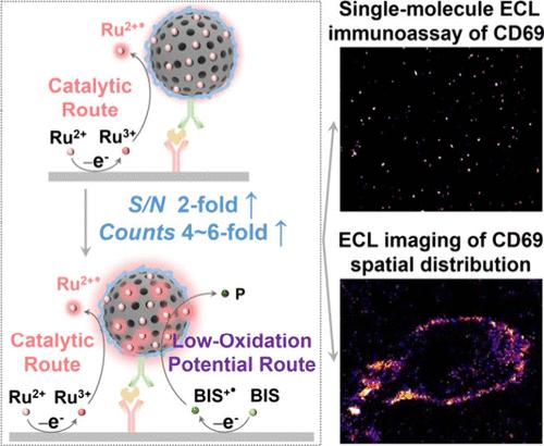
Immunotherapy using activated T cells as the weapon for controlling and eliminating tumor cells represents a promising therapeutic strategy for cancer treatment, in which accurate detection of T cell activation is critical for evaluating the immunotherapy efficacy. Activated T cells upregulate specific plasma membrane biomarkers, such as CD69, that can act as an indicator of activation status. In this work, we report a novel strategy of assessing T cell activation by electrochemiluminescence (ECL) imaging of CD69 expression on the plasma membrane. Mesoporous silica nanoparticles functionalized by polyethylenimine (PEI-SiO2) were prepared as both “nanocoreactant” and “nanoreactor” to remarkably enhance the ECL reaction through a dual-pathway scheme, in which both the catalytic route and the low-oxidation-potential route contribute to produce an enhanced ECL signal. Based on this strategy, subsequent conjugation of PEI-SiO2 with anti-CD69 antibodies allowed us to detect individual CD69 proteins by single-molecule ECL imaging, visualize their spatial distribution on the plasma membrane of activated T cell, and reveal their expression dynamics. The results establish a novel dark-field imaging strategy for assessing T cell activation, offering a low-background tool for investigating immune cell dynamics and immunotherapy efficacy through single-cell-level analysis of key biomarkers.
T细胞早期活化生物标志物的双途径增强单分子电化学发光成像
利用活化的T细胞作为控制和消灭肿瘤细胞的武器进行免疫治疗是一种很有前景的癌症治疗策略,其中准确检测T细胞活化是评估免疫治疗效果的关键。活化的T细胞上调特定的质膜生物标志物,如CD69,可以作为活化状态的一个指标。在这项工作中,我们报告了一种通过电化学发光(ECL)成像质膜上CD69表达来评估T细胞活化的新策略。制备了聚乙烯亚胺功能化介孔二氧化硅纳米颗粒(PEI-SiO2)作为“纳米反应物”和“纳米反应器”,通过催化途径和低氧化电位途径显著增强ECL反应信号。基于这一策略,随后将PEI-SiO2与抗CD69抗体偶联,使我们能够通过单分子ECL成像检测单个CD69蛋白,可视化其在活化T细胞质膜上的空间分布,并揭示其表达动态。研究结果建立了一种评估T细胞活化的新型暗场成像策略,通过单细胞水平的关键生物标志物分析,为研究免疫细胞动力学和免疫治疗疗效提供了低背景工具。
本文章由计算机程序翻译,如有差异,请以英文原文为准。
求助全文
约1分钟内获得全文
求助全文
来源期刊
Analytical Chemistry
化学-分析化学
CiteScore
12.10
自引率
12.20%
发文量
1949
审稿时长
1.4 months
期刊介绍:
Analytical Chemistry, a peer-reviewed research journal, focuses on disseminating new and original knowledge across all branches of analytical chemistry. Fundamental articles may explore general principles of chemical measurement science and need not directly address existing or potential analytical methodology. They can be entirely theoretical or report experimental results. Contributions may cover various phases of analytical operations, including sampling, bioanalysis, electrochemistry, mass spectrometry, microscale and nanoscale systems, environmental analysis, separations, spectroscopy, chemical reactions and selectivity, instrumentation, imaging, surface analysis, and data processing. Papers discussing known analytical methods should present a significant, original application of the method, a notable improvement, or results on an important analyte.
 求助内容:
求助内容: 应助结果提醒方式:


