Monitoring Chemotherapeutic Drugs Release from the Single Core–Shell Nanocarrier at a Monolayer Graphene-Based Total Internal Reflection Imaging Platform
{"title":"Monitoring Chemotherapeutic Drugs Release from the Single Core–Shell Nanocarrier at a Monolayer Graphene-Based Total Internal Reflection Imaging Platform","authors":"Jialu Chen, , , Zhimei He, , , Yapeng Li, , , Mengwei Chen, , , Zheng Cai, , , Yun Chen*, , , Yimin Fang*, , and , Hao Zhu*, ","doi":"10.1021/acs.analchem.5c04386","DOIUrl":null,"url":null,"abstract":"<p >Core–shell nanostructures have emerged as promising nanocarriers for diverse biomedical applications including thermal therapy, drug delivery, and tissue engineering. When core–shell nanocarriers are used in combination with chemotherapeutic drugs to achieve a synergistic effect, it is essential to monitor the release of chemotherapeutic drugs at the single nanoparticle level because of its inherent heterogeneity, which is also constructive for the rational design of relevant nanomedicine. Here, we developed a monolayer graphene-based total internal reflection imaging platform to study the release of chemotherapeutic drugs from the individual gold coated silica core–shell (SiO<sub>2</sub>@Au CS) nanoparticles by detecting their surface charges in real-time. The nanoparticles oscillate under an alternating electric field, and the oscillation signal was extracted from the intense background with a Fourier transform filter, enabling precise determination of the surface charges of the individual nanoparticles. The combination of monolayer graphene and an evanescent field ensures high sensitivity while suppressing the noise level of the method, which contributes to a superiorly low detection limit. With the present setup, the loading and release behavior of doxorubicin on the individual SiO<sub>2</sub>@Au CS nanoparticles were monitored, and the corresponding kinetic constants were extracted. In addition, the release rate of doxorubicin from the nanocomposites induced by the thermal effects were examined at the single nanoparticle level. This work offers a promising tool to study the heterogeneity of the nanocarriers’ surface charges to further reveal their structure–activity relationships, which will also provide guidance for the study of the mechanism of their effects <i>in vivo</i>.</p>","PeriodicalId":27,"journal":{"name":"Analytical Chemistry","volume":"97 40","pages":"22250–22258"},"PeriodicalIF":6.7000,"publicationDate":"2025-10-05","publicationTypes":"Journal Article","fieldsOfStudy":null,"isOpenAccess":false,"openAccessPdf":"","citationCount":"0","resultStr":null,"platform":"Semanticscholar","paperid":null,"PeriodicalName":"Analytical Chemistry","FirstCategoryId":"92","ListUrlMain":"https://pubs.acs.org/doi/10.1021/acs.analchem.5c04386","RegionNum":1,"RegionCategory":"化学","ArticlePicture":[],"TitleCN":null,"AbstractTextCN":null,"PMCID":null,"EPubDate":"","PubModel":"","JCR":"Q1","JCRName":"CHEMISTRY, ANALYTICAL","Score":null,"Total":0}
引用次数: 0
Abstract
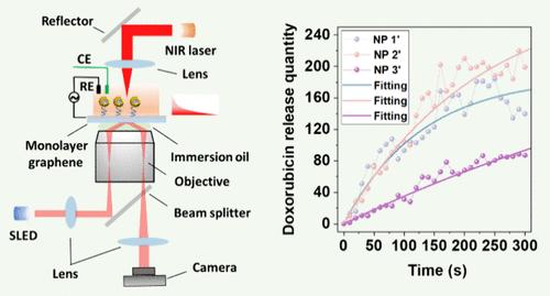
Core–shell nanostructures have emerged as promising nanocarriers for diverse biomedical applications including thermal therapy, drug delivery, and tissue engineering. When core–shell nanocarriers are used in combination with chemotherapeutic drugs to achieve a synergistic effect, it is essential to monitor the release of chemotherapeutic drugs at the single nanoparticle level because of its inherent heterogeneity, which is also constructive for the rational design of relevant nanomedicine. Here, we developed a monolayer graphene-based total internal reflection imaging platform to study the release of chemotherapeutic drugs from the individual gold coated silica core–shell (SiO2@Au CS) nanoparticles by detecting their surface charges in real-time. The nanoparticles oscillate under an alternating electric field, and the oscillation signal was extracted from the intense background with a Fourier transform filter, enabling precise determination of the surface charges of the individual nanoparticles. The combination of monolayer graphene and an evanescent field ensures high sensitivity while suppressing the noise level of the method, which contributes to a superiorly low detection limit. With the present setup, the loading and release behavior of doxorubicin on the individual SiO2@Au CS nanoparticles were monitored, and the corresponding kinetic constants were extracted. In addition, the release rate of doxorubicin from the nanocomposites induced by the thermal effects were examined at the single nanoparticle level. This work offers a promising tool to study the heterogeneity of the nanocarriers’ surface charges to further reveal their structure–activity relationships, which will also provide guidance for the study of the mechanism of their effects in vivo.
期刊介绍:
Analytical Chemistry, a peer-reviewed research journal, focuses on disseminating new and original knowledge across all branches of analytical chemistry. Fundamental articles may explore general principles of chemical measurement science and need not directly address existing or potential analytical methodology. They can be entirely theoretical or report experimental results. Contributions may cover various phases of analytical operations, including sampling, bioanalysis, electrochemistry, mass spectrometry, microscale and nanoscale systems, environmental analysis, separations, spectroscopy, chemical reactions and selectivity, instrumentation, imaging, surface analysis, and data processing. Papers discussing known analytical methods should present a significant, original application of the method, a notable improvement, or results on an important analyte.
 求助内容:
求助内容: 应助结果提醒方式:


