Eleni Sinopoulou, Michelle W Chow, Numaira Obaid, Emily Chong, Yvette S Nout-Lomas, Rachele Wurr, Ryan Macon, J Russell Huie, Adam R Ferguson, Mark H Tuszynski, Michael S Beattie, Jacqueline C Bresnahan, Carolyn J Sparrey
{"title":"Spinal Cord Injury in Real Time: Intra-Operative Ultrasound for Acute Phase Examination in Non-Human Primates.","authors":"Eleni Sinopoulou, Michelle W Chow, Numaira Obaid, Emily Chong, Yvette S Nout-Lomas, Rachele Wurr, Ryan Macon, J Russell Huie, Adam R Ferguson, Mark H Tuszynski, Michael S Beattie, Jacqueline C Bresnahan, Carolyn J Sparrey","doi":"10.3390/brainsci15091005","DOIUrl":null,"url":null,"abstract":"<p><strong>Background: </strong>A spinal cord contusion injury is among the most clinically relevant models for studying pathophysiology and for developing potential therapeutic interventions for spinal cord injuries (SCI).</p><p><strong>Methods: </strong>In this study, we implemented an intra-operative ultrasound (IOU) approach to precisely locate and examine the lesion site at 5 and 10 min post-injury after a cervical hemi-contusion injury in a non-human primate (NHP) model. We assessed acute lesion progression from 5 to 10 min and then compared that to the lesion extent as measured by MRI 3 weeks later.</p><p><strong>Results: </strong>We observed a small increase in the rostrocaudal and mediolateral lesion area (mm<sup>2</sup>) from 5 to 10 min and a further 26% increase in the mediolateral lesion extent when comparing 5 and 10 min to 3 weeks post-injury.</p><p><strong>Conclusions: </strong>By enabling high-resolution ultrasound visualization of the hemicontusion lesion in vivo, this approach can provide critical insights into the early progression of SCI. It can help with further refining this preclinical SCI model and provide significant predictive value for the animals' recovery post-injury.</p>","PeriodicalId":9095,"journal":{"name":"Brain Sciences","volume":"15 9","pages":""},"PeriodicalIF":2.8000,"publicationDate":"2025-09-17","publicationTypes":"Journal Article","fieldsOfStudy":null,"isOpenAccess":false,"openAccessPdf":"https://www.ncbi.nlm.nih.gov/pmc/articles/PMC12468102/pdf/","citationCount":"0","resultStr":null,"platform":"Semanticscholar","paperid":null,"PeriodicalName":"Brain Sciences","FirstCategoryId":"3","ListUrlMain":"https://doi.org/10.3390/brainsci15091005","RegionNum":3,"RegionCategory":"医学","ArticlePicture":[],"TitleCN":null,"AbstractTextCN":null,"PMCID":null,"EPubDate":"","PubModel":"","JCR":"Q3","JCRName":"NEUROSCIENCES","Score":null,"Total":0}
引用次数: 0
Abstract
Background: A spinal cord contusion injury is among the most clinically relevant models for studying pathophysiology and for developing potential therapeutic interventions for spinal cord injuries (SCI).
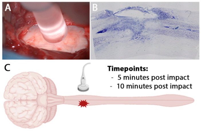
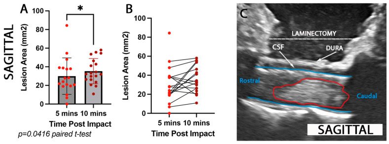
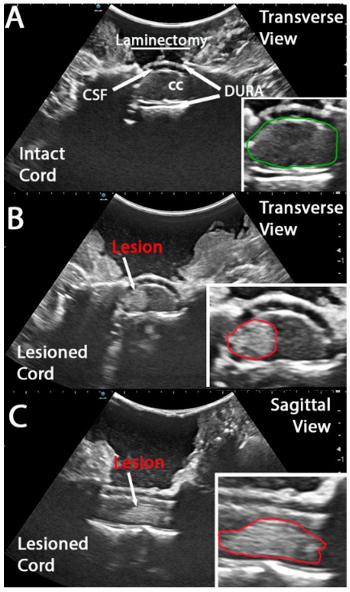
Methods: In this study, we implemented an intra-operative ultrasound (IOU) approach to precisely locate and examine the lesion site at 5 and 10 min post-injury after a cervical hemi-contusion injury in a non-human primate (NHP) model. We assessed acute lesion progression from 5 to 10 min and then compared that to the lesion extent as measured by MRI 3 weeks later.
Results: We observed a small increase in the rostrocaudal and mediolateral lesion area (mm2) from 5 to 10 min and a further 26% increase in the mediolateral lesion extent when comparing 5 and 10 min to 3 weeks post-injury.
Conclusions: By enabling high-resolution ultrasound visualization of the hemicontusion lesion in vivo, this approach can provide critical insights into the early progression of SCI. It can help with further refining this preclinical SCI model and provide significant predictive value for the animals' recovery post-injury.
期刊介绍:
Brain Sciences (ISSN 2076-3425) is a peer-reviewed scientific journal that publishes original articles, critical reviews, research notes and short communications in the areas of cognitive neuroscience, developmental neuroscience, molecular and cellular neuroscience, neural engineering, neuroimaging, neurolinguistics, neuropathy, systems neuroscience, and theoretical and computational neuroscience. Our aim is to encourage scientists to publish their experimental and theoretical results in as much detail as possible. There is no restriction on the length of the papers. The full experimental details must be provided so that the results can be reproduced. Electronic files or software regarding the full details of the calculation and experimental procedure, if unable to be published in a normal way, can be deposited as supplementary material.
 求助内容:
求助内容: 应助结果提醒方式:


