Enhanced detection of PSA by nanoscale plasmonic devices and Raman spectroscopy
IF 3.1
Q2 ENGINEERING, ELECTRICAL & ELECTRONIC
引用次数: 0
Abstract
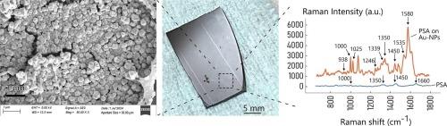
Prostate-specific antigen (PSA) is a crucial biomarker for the early detection and monitoring of prostate cancer (PC). In this study, we present a biosensing approach that integrates plasmonic nanostructures with surface-enhanced Raman spectroscopy (SERS) for the ultrasensitive detection of PSA in diluted solution. Our sensor device consists of ordered arrays of densely packed gold nanoparticles (Au NPs), fabricated using a combination of optical-lithography and electroless deposition techniques. The plasmonic properties of the Au NPs enhance the Raman scattering effect, significantly improving sensitivity and detection limits. We demonstrate the device's capability to detect PSA at vanishingly low concentrations – as low as - well below the 4 ng/mL threshold used in clinical practice. Data analysis of Raman spectra illustrate that the response of the sensor device to PSA exhibits two distinct, approximately linear regimes. In the first regime (I), the Raman intensity increases with PSA concentration. In the second regime (II), the intensity decreases as concentration continues to rise. The transition between these regimes occurs at around . The existence of these regimes is explained by the peculiar behavior of surface enhanced Raman substrates, where the signal intensity non-linearly depends on the distance from the active metal nano-surface. At higher PSA concentrations, the biomarker may accumulate on the Au NPs, hampering the efficiency of sensing. These findings suggest that this plasmonic-SERS platform could provide a highly effective, non-invasive tool for PSA detection, potentially improving PC diagnostics.
利用纳米等离子体器件和拉曼光谱增强PSA检测
前列腺特异性抗原(PSA)是早期发现和监测前列腺癌的重要生物标志物。在这项研究中,我们提出了一种生物传感方法,将等离子体纳米结构与表面增强拉曼光谱(SERS)相结合,用于稀释溶液中PSA的超灵敏检测。我们的传感器装置由有序排列的密集排列的金纳米颗粒(Au NPs)组成,采用光学光刻和化学沉积技术相结合的方式制造。金纳米粒子的等离子体特性增强了拉曼散射效应,显著提高了灵敏度和检测限。我们展示了该设备在极低浓度下检测PSA的能力-低至38pg/mL -远低于临床实践中使用的4ng /mL阈值。拉曼光谱的数据分析表明,传感器器件对PSA的响应表现出两种不同的近似线性状态。在第一阶段(I),拉曼强度随PSA浓度的增加而增加。在第二种状态(II)中,浓度继续升高,强度降低。这些状态之间的转变发生在3ng/mL左右。这些机制的存在可以用表面增强拉曼衬底的特殊行为来解释,其中信号强度非线性地依赖于与活性金属纳米表面的距离。在较高的PSA浓度下,生物标志物可能积聚在Au NPs上,阻碍了传感的效率。这些发现表明,这种等离子体- sers平台可以为PSA检测提供一种高效、无创的工具,有可能改善PC诊断。
本文章由计算机程序翻译,如有差异,请以英文原文为准。
求助全文
约1分钟内获得全文
求助全文
来源期刊
Micro and Nano Engineering
Engineering-Electrical and Electronic Engineering
CiteScore
3.30
自引率
0.00%
发文量
67
审稿时长
80 days
 求助内容:
求助内容: 应助结果提醒方式:


