Satish K Gupta, Andrew Carkeet, Scott A Read, Stephen J Vincent, David A Atchison
{"title":"Contribution and Compensation Effects of Refracting Components to Ocular Aberrations in Keratoconus.","authors":"Satish K Gupta, Andrew Carkeet, Scott A Read, Stephen J Vincent, David A Atchison","doi":"10.1167/iovs.66.11.64","DOIUrl":null,"url":null,"abstract":"<p><strong>Purpose: </strong>The purpose of this study was to determine the (i) contributions of refracting components to ocular aberrations and (ii) compensation effects exhibited by these components in keratoconus.</p><p><strong>Methods: </strong>Right eyes of 14 keratoconus and 20 control participants were analyzed using 5 mm pupils. Ocular aberrations were measured with a Hartmann-Shack aberrometer. Corneas were imaged with a Scheimpflug tomographer. Three-dimensional models of the total cornea and anterior cornea were created. Raytracing included correct object-image conjugates and corneal decentration relative to the aberrometer pupillary center to determine the total corneal and anterior corneal aberrations. Posterior corneal and lenticular aberrations were computed. Compensation effects (%) were calculated: 100 (anterior corneal-total corneal aberration)/anterior corneal aberration, and 100 (total corneal-ocular aberration)/total corneal aberration.</p><p><strong>Results: </strong>Considering coefficients for the total cornea with absolute values >0.05 µm, for both corneal surfaces, keratoconus had higher magnitudes than controls for C(2,-2), C(3,-3), C(3,-1), C(4,-2), total root mean square (RMS), higher-order RMS (HORMS), and J45. Both surfaces' RMS aberrations were approximately 2 to 5 times higher in keratoconus than in controls. Anterior corneal RMS aberrations were approximately 5 times (keratoconus) and approximately 3 to 4 times (controls) higher than those of the posterior cornea. Posterior corneal compensations for anterior corneal aberrations were higher in keratoconus than in controls for C(3,-3) (21%, decompensation of -14%), C(3,-1) (21%, -33%), C(4,-2) (27%, -10%), C(4,+2) (22%, 10%), HORMS (20%, 2%), and J0 (68%, 66%), as were lenticular compensations for total corneal aberrations for C(2,-2) (40%, -64%), C(2,+2) (70%, 60%), total RMS (21%, 20%), and J0 (642%, -55%).</p><p><strong>Conclusions: </strong>Keratoconic eyes exhibited higher anterior and posterior corneal aberrations than control eyes. The posterior cornea and lens compensated partly for the anterior cornea and total cornea, respectively, with greater percentage compensations in keratoconus.</p>","PeriodicalId":14620,"journal":{"name":"Investigative ophthalmology & visual science","volume":"66 11","pages":"64"},"PeriodicalIF":4.7000,"publicationDate":"2025-08-01","publicationTypes":"Journal Article","fieldsOfStudy":null,"isOpenAccess":false,"openAccessPdf":"https://www.ncbi.nlm.nih.gov/pmc/articles/PMC12393118/pdf/","citationCount":"0","resultStr":null,"platform":"Semanticscholar","paperid":null,"PeriodicalName":"Investigative ophthalmology & visual science","FirstCategoryId":"3","ListUrlMain":"https://doi.org/10.1167/iovs.66.11.64","RegionNum":2,"RegionCategory":"医学","ArticlePicture":[],"TitleCN":null,"AbstractTextCN":null,"PMCID":null,"EPubDate":"","PubModel":"","JCR":"Q1","JCRName":"OPHTHALMOLOGY","Score":null,"Total":0}
引用次数: 0
Abstract
Purpose: The purpose of this study was to determine the (i) contributions of refracting components to ocular aberrations and (ii) compensation effects exhibited by these components in keratoconus.
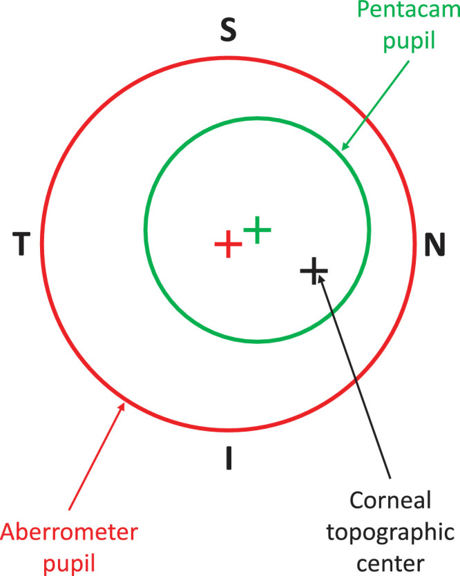
Methods: Right eyes of 14 keratoconus and 20 control participants were analyzed using 5 mm pupils. Ocular aberrations were measured with a Hartmann-Shack aberrometer. Corneas were imaged with a Scheimpflug tomographer. Three-dimensional models of the total cornea and anterior cornea were created. Raytracing included correct object-image conjugates and corneal decentration relative to the aberrometer pupillary center to determine the total corneal and anterior corneal aberrations. Posterior corneal and lenticular aberrations were computed. Compensation effects (%) were calculated: 100 (anterior corneal-total corneal aberration)/anterior corneal aberration, and 100 (total corneal-ocular aberration)/total corneal aberration.
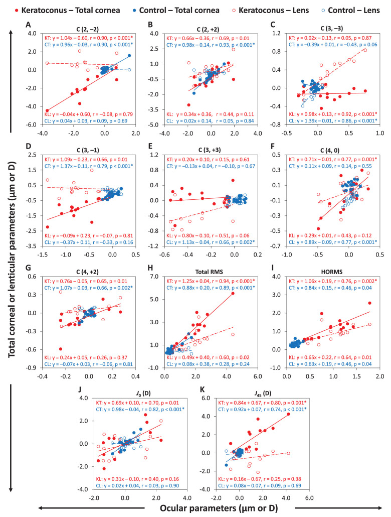
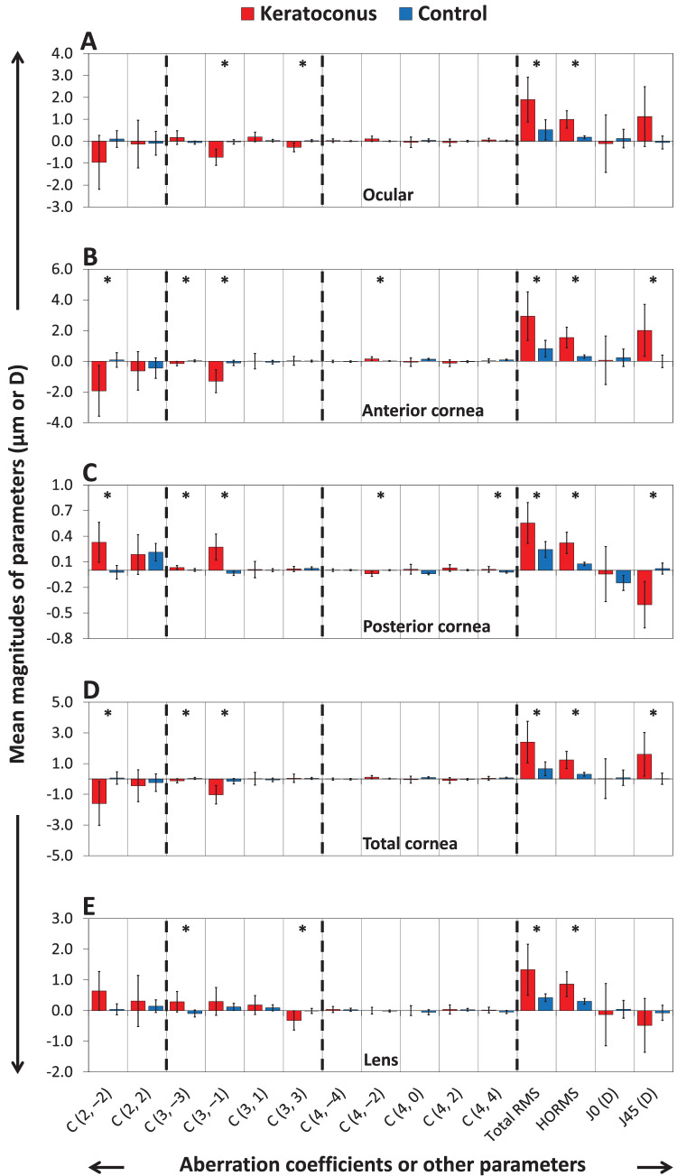
Results: Considering coefficients for the total cornea with absolute values >0.05 µm, for both corneal surfaces, keratoconus had higher magnitudes than controls for C(2,-2), C(3,-3), C(3,-1), C(4,-2), total root mean square (RMS), higher-order RMS (HORMS), and J45. Both surfaces' RMS aberrations were approximately 2 to 5 times higher in keratoconus than in controls. Anterior corneal RMS aberrations were approximately 5 times (keratoconus) and approximately 3 to 4 times (controls) higher than those of the posterior cornea. Posterior corneal compensations for anterior corneal aberrations were higher in keratoconus than in controls for C(3,-3) (21%, decompensation of -14%), C(3,-1) (21%, -33%), C(4,-2) (27%, -10%), C(4,+2) (22%, 10%), HORMS (20%, 2%), and J0 (68%, 66%), as were lenticular compensations for total corneal aberrations for C(2,-2) (40%, -64%), C(2,+2) (70%, 60%), total RMS (21%, 20%), and J0 (642%, -55%).
Conclusions: Keratoconic eyes exhibited higher anterior and posterior corneal aberrations than control eyes. The posterior cornea and lens compensated partly for the anterior cornea and total cornea, respectively, with greater percentage compensations in keratoconus.
期刊介绍:
Investigative Ophthalmology & Visual Science (IOVS), published as ready online, is a peer-reviewed academic journal of the Association for Research in Vision and Ophthalmology (ARVO). IOVS features original research, mostly pertaining to clinical and laboratory ophthalmology and vision research in general.

 求助内容:
求助内容: 应助结果提醒方式:


