{"title":"Functional Anatomy of Ocular Counter-Rolling in Superior Oblique Palsy.","authors":"Joseph L Demer, Robert A Clark","doi":"10.1167/iovs.66.12.6","DOIUrl":null,"url":null,"abstract":"<p><strong>Purpose: </strong>Simulations suggest that displacement of rectus extraocular muscle pulleys in superior oblique (SO) palsy accounts for incomitant strabismus patterns even without postulating SO contractile weakness. We asked how rectus extraocular muscle pulleys reorient during head tilt in SO palsy.</p><p><strong>Methods: </strong>In 13 subjects with unilateral SO palsy, supine magnetic resonance imaging (MRI) in 2-mm-thick quasi-coronal planes in target-controlled central gaze was repeated in both lateral decubitus positions equivalent to 90° head tilts. From extraocular muscle centroids, we computed oculocentric pulley coordinates and compartmental posterior partial volumes (PPVs) of the rectus and SO muscles.</p><p><strong>Results: </strong>Validating atrophy, PPV of the palsied SO was smaller than its fellow (P < 10-4). In fellow orbits, the array of all four rectus pulleys exhibited counter-rotation during head tilt (P < 0.03), averaging 5.9°. The palsied pulley array was in both tilts excyclorotated relative to the fellow orbit, particularly by 4° to 5° for horizontal rectus pulleys (P < 0.03), and also counter-rotated with head tilt similarly to the fellow orbit. Differential compartmental changes in PPV were significant in the lateral and superior rectus and SO muscles of the fellow orbit that were consistent with observed torsion, but were absent in the palsied orbit.</p><p><strong>Conclusions: </strong>Similar counter-rotation of the rectus pulley array during head tilt occurs in both eyes in unilateral SO palsy, but superimposed on excyclorotation of the array in the palsied orbit. Differential compartmental change in PPV occurs during head tilt in the lateral and superior rectus muscles of the fellow but not palsied orbit and could augment ocular counter-rolling.</p>","PeriodicalId":14620,"journal":{"name":"Investigative ophthalmology & visual science","volume":"66 12","pages":"6"},"PeriodicalIF":4.7000,"publicationDate":"2025-09-02","publicationTypes":"Journal Article","fieldsOfStudy":null,"isOpenAccess":false,"openAccessPdf":"https://www.ncbi.nlm.nih.gov/pmc/articles/PMC12410258/pdf/","citationCount":"0","resultStr":null,"platform":"Semanticscholar","paperid":null,"PeriodicalName":"Investigative ophthalmology & visual science","FirstCategoryId":"3","ListUrlMain":"https://doi.org/10.1167/iovs.66.12.6","RegionNum":2,"RegionCategory":"医学","ArticlePicture":[],"TitleCN":null,"AbstractTextCN":null,"PMCID":null,"EPubDate":"","PubModel":"","JCR":"Q1","JCRName":"OPHTHALMOLOGY","Score":null,"Total":0}
引用次数: 0
Abstract
Purpose: Simulations suggest that displacement of rectus extraocular muscle pulleys in superior oblique (SO) palsy accounts for incomitant strabismus patterns even without postulating SO contractile weakness. We asked how rectus extraocular muscle pulleys reorient during head tilt in SO palsy.
Methods: In 13 subjects with unilateral SO palsy, supine magnetic resonance imaging (MRI) in 2-mm-thick quasi-coronal planes in target-controlled central gaze was repeated in both lateral decubitus positions equivalent to 90° head tilts. From extraocular muscle centroids, we computed oculocentric pulley coordinates and compartmental posterior partial volumes (PPVs) of the rectus and SO muscles.
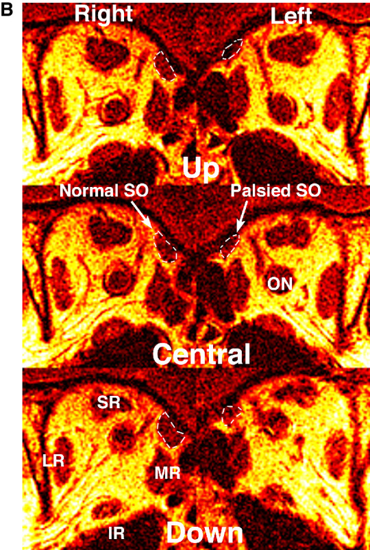
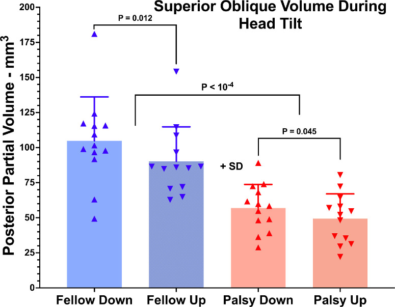
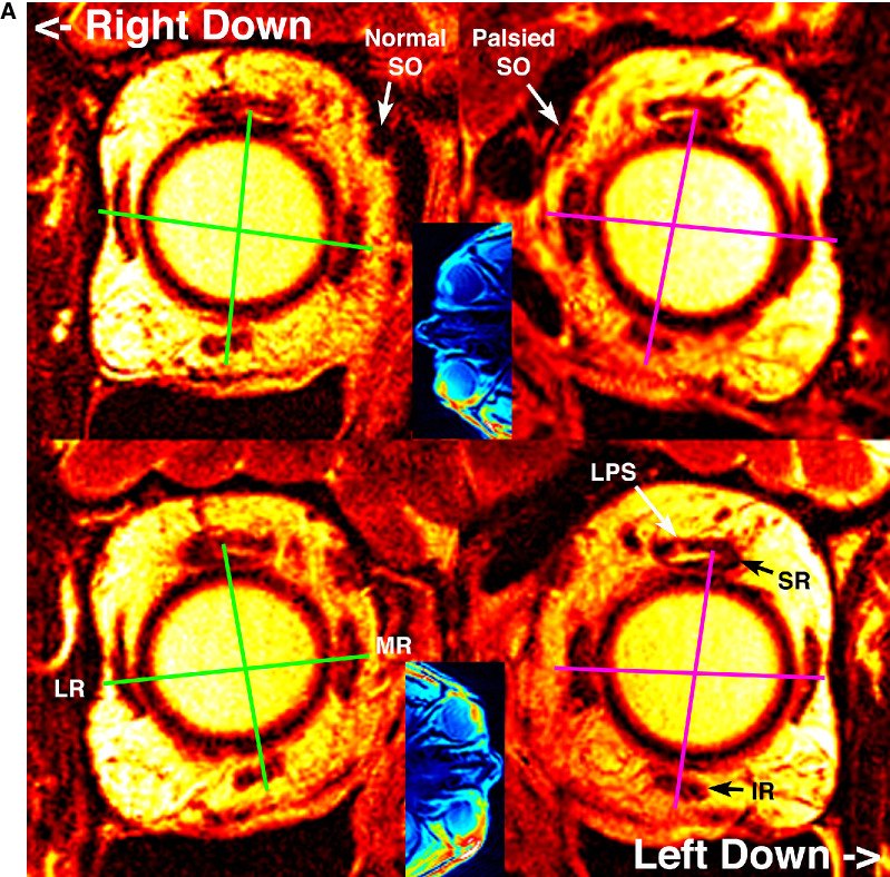
Results: Validating atrophy, PPV of the palsied SO was smaller than its fellow (P < 10-4). In fellow orbits, the array of all four rectus pulleys exhibited counter-rotation during head tilt (P < 0.03), averaging 5.9°. The palsied pulley array was in both tilts excyclorotated relative to the fellow orbit, particularly by 4° to 5° for horizontal rectus pulleys (P < 0.03), and also counter-rotated with head tilt similarly to the fellow orbit. Differential compartmental changes in PPV were significant in the lateral and superior rectus and SO muscles of the fellow orbit that were consistent with observed torsion, but were absent in the palsied orbit.
Conclusions: Similar counter-rotation of the rectus pulley array during head tilt occurs in both eyes in unilateral SO palsy, but superimposed on excyclorotation of the array in the palsied orbit. Differential compartmental change in PPV occurs during head tilt in the lateral and superior rectus muscles of the fellow but not palsied orbit and could augment ocular counter-rolling.
期刊介绍:
Investigative Ophthalmology & Visual Science (IOVS), published as ready online, is a peer-reviewed academic journal of the Association for Research in Vision and Ophthalmology (ARVO). IOVS features original research, mostly pertaining to clinical and laboratory ophthalmology and vision research in general.

 求助内容:
求助内容: 应助结果提醒方式:


