Robust cortical thickness estimation in the presence of partial volumes using adaptive diffusion equation
IF 2.3
4区 医学
Q2 BIOCHEMICAL RESEARCH METHODS
引用次数: 0
Abstract
Background:
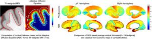
Automated estimation of cortical thickness in brain MRI is a critical step when investigating neuroanatomical population differences and changes associated with normal development and aging, as well as in neurodegenerative diseases such as Alzheimer’s and Parkinson’s. The limited spatial resolution of the scanner leads to partial volume effects, where each voxel in the scanned image may represent a mixture of more than one type of tissue. Due to the highly convoluted structure of the cortex, this can have a significant impact on the accuracy of thickness estimates, particularly if a hard intensity threshold is used to delineate cortical boundaries.
New methods:
In this paper, we describe a novel method based on an adaptive diffusion equation (ADE) that explicitly accounts for the presence of partial tissue volumes to estimate cortical thickness more accurately. The diffusivity term uses gray matter fractions to incorporate partial tissue volumes into the thickness calculation.
Results:
We show that the proposed method is robust to the effects of finite voxel resolution and blurring. The method was validated through simulations, comparisons with histological measurements reported in the literature, and single- and multi-scanner test–retest studies.
Comparison with existing methods
: The proposed method was compared with methods based on the Laplace equation, a linked distance metric, and the FreeSurfer software package.
Conclusion:
We introduced a novel method (ADE) for estimating cortical thickness that is robust to variations in image resolution and scanner field strength. ADE yields accurate, histologically consistent thickness estimates and demonstrates superior consistency in multi-scanner test–retest studies.
基于自适应扩散方程的部分体积存在下的鲁棒皮质厚度估计
背景:在研究与正常发育和衰老相关的神经解剖学群体差异和变化以及阿尔茨海默病和帕金森病等神经退行性疾病时,脑MRI中皮质厚度的自动估计是一个关键步骤。扫描仪的有限空间分辨率导致部分体积效应,其中扫描图像中的每个体素可能代表一种以上组织类型的混合物。由于皮质的高度卷曲结构,这可能对厚度估计的准确性产生重大影响,特别是如果使用硬强度阈值来划定皮质边界。新方法:在本文中,我们描述了一种基于自适应扩散方程(ADE)的新方法,该方法明确地考虑了部分组织体积的存在,以更准确地估计皮层厚度。扩散率项使用灰质分数将部分组织体积合并到厚度计算中。结果:该方法对有限体素分辨率和模糊的影响具有较强的鲁棒性。该方法通过模拟、与文献中报道的组织学测量结果的比较以及单扫描仪和多扫描仪测试-重测试研究进行了验证。与现有方法的比较:将本文方法与基于拉普拉斯方程、链接距离度量和FreeSurfer软件包的方法进行了比较。结论:我们引入了一种新的方法(ADE)来估计皮质厚度,该方法对图像分辨率和扫描仪场强的变化具有鲁棒性。ADE产生准确的,组织学上一致的厚度估计,并在多扫描仪测试-重测研究中显示出优越的一致性。
本文章由计算机程序翻译,如有差异,请以英文原文为准。
求助全文
约1分钟内获得全文
求助全文
来源期刊

Journal of Neuroscience Methods
医学-神经科学
CiteScore
7.10
自引率
3.30%
发文量
226
审稿时长
52 days
期刊介绍:
The Journal of Neuroscience Methods publishes papers that describe new methods that are specifically for neuroscience research conducted in invertebrates, vertebrates or in man. Major methodological improvements or important refinements of established neuroscience methods are also considered for publication. The Journal''s Scope includes all aspects of contemporary neuroscience research, including anatomical, behavioural, biochemical, cellular, computational, molecular, invasive and non-invasive imaging, optogenetic, and physiological research investigations.
 求助内容:
求助内容: 应助结果提醒方式:


